Please enter url.
Login
Logout
Please enter url.
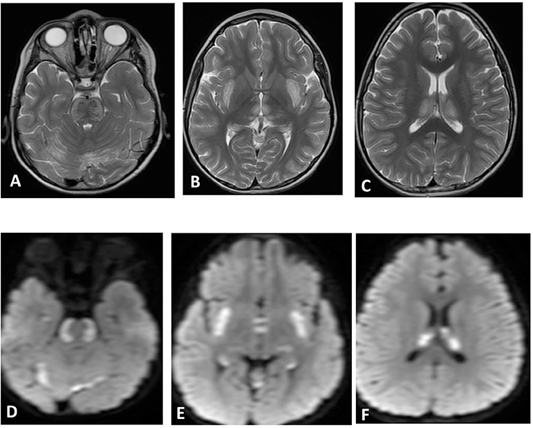
| Brain MRI. Axial T2 (A-C) and diffusion (D-F) showing swelling and ...
researchgate.net
source
Comments
(a) Sagittal CT image in an 11-year-old male shows a pineal mass that ...
Frontiers | Acute Necrotizing Encephalopathy of Childhood: A ...
Radiological findings for patients with supratentorial infarcts. We ...
MRI brain. (A) Diffusion weighted image-revealing restriction of ...
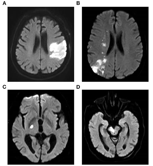
Cerebral Venous Thrombosis. A 28-year-old female was presented with ...
Representative brain MRI acquired 4 days after symptom onset in a male ...
Patient 1: facial features with down-slanting palpebral fissures ...
MRI of Patient 2, revealing multiple lacunar infarctions of frontal ...
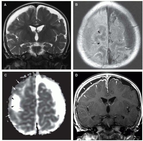
Axial T2 weighted image showing supra- and infra-tentorial high signals ...
Infections of the Developing and Mature Nervous System | Radiology Key
MRI demonstrating interval improvement immediately following repeat ...
Brain MRI in the 7th (a–c) and 9th (d–f) week after admission. FLAIR ...
Malik GHANNAM | Vascular Neurology Fellow | MBBCh | University of Iowa ...
Cerebellitis. A 19-year-old primigravida presented 4 days after labor ...
Cureus | Posterior Reversible Encephalopathy Syndrome With Hemorrhagic ...
Frontiers | Acute Necrotizing Encephalopathy of Childhood: A ...
| Hyperintense vessel sign (HVS): (A) Score of 1, restricted to Sylvian ...
High-resolution, T2-weighted, FSE MRI (voxels of 0.47 mm ϫ 0.47 mm ϫ 2 ...
Early Subacute Hematoma - Cytotoxic Edema - Mussen Healthcare
(PDF) Bilateral Thalamic Infarction and DSA Demonstrated AOP after ...
Biotin and Thiamine Responsive Basal Ganglia Disease – A vital ...
Representative images of infarcts in multiple cerebrovascular ...
Artifacts secondary to the shunt reservoir. The SS-GRE MR image is the ...
A, Axial CT scan shows a mass lesion in the trigone of the left lateral ...
Frontiers | Clinical Utility of the Serum Level of Lipoprotein-Related ...
Encephalitis and Thalamic Injury From Neuroinvasive West Nile Virus in ...
Nucleus Subthalamicus - Magnetic Resonance - 78 Steps Health
A 53-year-old man with hyperacute intracerebral hematoma seen on MR ...
Radiographic images of brain disorders associated with Van Der Knaap ...
Herpes Simplex Virus Type-1 Encephalitis and Occipital Ischemic Stroke ...
Hemiballism with leg predominance caused by contralateral subthalamic ...
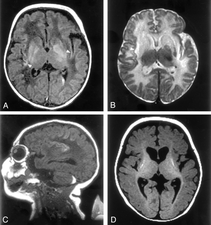
Cerebral MRI abnormalities associated with vigabatrin therapy - Pearl ...
Axial T2 ( ) and fluid-attenuated inversion recovery ( ) images show ...
Analysis of Cystic Intracranial Lesions Performed with Fluid-Attenuated ...
Brain MR Imaging in Neonatal Hyperammonemic Encephalopathy Resulting ...
Lesions-On-Brain-MRI
MS-Brain-MRI-with-Contrast
Lesions-On-MRI-Brain-Scan
Lymphoma-Brain-MRI
Brain-MRI-Showing-Lesions
MRI-Brain-Cerebellum
PML-MRI-Brain
Meningitis-MRI-Brain
MRI-of-Brain-After-Stroke
Brain-Infection-MRI
Fibromyalgia-Brain-MRI
Axial-Brain-MRI
Abnormal-MRI-Brain-Lesions
MRI-Brain-Signs
Brain-MRI-of-MS-Patients
Brain-Stem-MRI-Images