Please enter url.
Login
Logout
Please enter url.
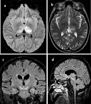
Representative brain MRI acquired 4 days after symptom onset in a male ...
researchgate.net
source
Comments
Representative head CT a image after 8 days of acyclovir treatment ...
West Nile virus encephalitis outbreak in the Guadalquivir river basin ...
Early HSV-1 encephalitis. Hyperintense lesions involving enlarged left ...
HSV-2 encephalitis. Sequential axial FLAIR images ( A – D ) show patchy ...
Vigabatrin‐associated reversible MRI signal changes in patients with ...
Reversible movement disorders due to toxoplasmosis as initial ...
Magnetic Resonance Imaging Showing Left Occipital Lobe Lymphoma Before ...
Infections of the Developing and Mature Nervous System | Radiology Key
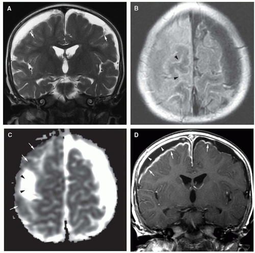
Representative images of infarcts in multiple cerebrovascular ...
Measuring signal intensities on diffusion-weighted imaging (DWI) and ...
Brain (A) T2WI (B) FLAIR (C) DIR (D) FLAIR and (E) DIR. FLAIR ...
Patient 2. A 48-year-old man with fulminant acute hepatic failure due ...
Analysis of Cystic Intracranial Lesions Performed with Fluid-Attenuated ...
Pathology of human organic mercury poisoning: Lessons from an autopsy ...
Acute encephalitis with refractory, repetitive partial seizures - Brain ...
Indications and Imaging Modality of Choice in Pediatric Headache ...
Cerebral toxoplasmosis responding to therapy. (A) Axial T2-weighted ...
Disseminated lesions in 3 patients demonstrated at contrast-enhanced MR ...
Analysis of Cystic Intracranial Lesions Performed with Fluid-Attenuated ...
Degenerative Disorders of the Newborn | Neupsy Key
Brain MRI images of the patient in the different phases of the disease ...
Pneumocephalus Mimicking Cerebral Cavernous Malformations in MR ...
The intraventricular DWI hyperintense scolex is appreciated inside a ...
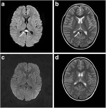
Clinically mild encephalitis/encephalopathy with a reversible splenial ...
Acute cortical blindness caused by neuropsychiatric systemic lupus ...
a DWI with intratumoral bleeding in a GBM patient without AT. b ...
Hemiballism with leg predominance caused by contralateral subthalamic ...
Magnetic Resonance Imaging (MRI) of the Brain on Hospital Admission ...
The usefulness of values of the diffusion-weighted imaging (DWI) ratio ...
MRI appearance of white matter changes in axial sections of patients ...
MR images obtained pre and postoperatively represent ischemic lesions ...
(A and B) DWI shows a 1-cm right convexity hyperintense mass (white ...
Asymptomatic Large T2 High-Signal Pontine Lesions that Are Different ...
Limbic Encephalitis Associated With Anti–Voltage-Gated Potassium ...
Magnetic resonance images. (A) T2 axial and (B) T1 axial multiplanar ...
Encephalitis-On-MRI
Herpes-Viral-Encephalitis
Viral-Encephalitis-MRI
Encephalitis-MRI-Brain
CMV-Encephalitis
HSV-MRI
Acute-Encephalitis
Brainstem-Encephalitis
HSV-Encephalitis-MRI-Findings
Encephalitis-Brain-Scan
Japanese-Encephalitis-MRI
Temporal-Lobe-Encephalitis
HSV-Encephalitis-CT-Head
Autoimmune-Encephalitis-MRI
HSV-Encephalitis-CSF
HSV-Retina-MRI-Brain