Please enter url.
Login
Logout
Please enter url.
MRI demonstrating interval improvement immediately following repeat ...
researchgate.net
source
Comments
MRI demonstrating interval improvement immediately following repeat ...
Central Nervous System Infection | Radiology Key
The intraventricular DWI hyperintense scolex is appreciated inside a ...
Susceptibility-Weighted Imaging in Pediatric Arterial Ischemic Stroke ...
MRI results for a 13-year-old-boy with SLI a) axial FSE T2 ...
The brain MRI changes in Fabry disease. A DWI sequence showed recent ...
Fluorescence-guided surgery for glioblastoma multiforme using high-dose ...
AIDS-related cerebral toxoplasmosis with hyperintense foci on T1 ...
Location of DAI lesions in the deep intra-axial structures. A and B ...
Early-onset infant epileptic encephalopathy associated with a de novo ...
Parasitic Diseases of the Central Nervous System | Radiology Key
Morphological and functional MRI, MRS, perfusion and diffusion changes ...
Magnetic resonance imaging in Case 4 show a right perisylvian mass ...
ADC Values in myeloid sarcoma and normal brain | Download Table
Typical diagnostic procedures findings. a Brain MRI. Sagittal and axial ...
Left parietal high grade glioma, GBM, (WHO grade IV) in a 55-year-old ...
Axial MR images demonstrating diffusion restriction (A & B ...
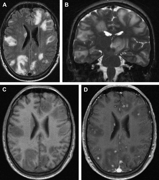
(PDF) Rituximab-Associated Progressive Multifocal Leukoencephalopathy ...
Follow-up MRI 3 days after the initial MRI, coronal FLAIR ( A and B ...
Case 1. A: T 2 -weighted magnetic resonance (MR) image showing an ...
PXA of the right temporal lobe. The lesion shows typical... | Download ...
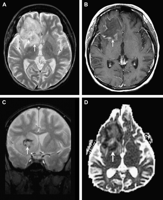
Nocardia farcinica brain abscess in an immunocompetent patient treated ...
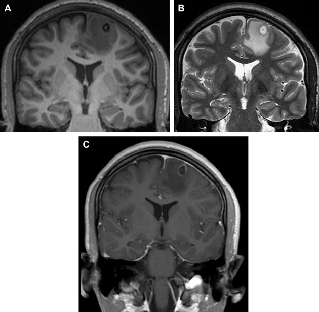
Concomitant parasagittal meningioma and adjacent intracranial abscess ...
Incidental Cerebral Toxocarosis as Confirmed by Cerebrospinal Fluid ...
Choreiform movements and basal ganglia calcification as a presentation ...
Prenatal Neurological Diagnosis: Challenges in Neuroimaging, Prognostic ...
Neuroimaging in the Brain in HIV-1–Infected Patients | Radiology Key
a and b: Axial, FLAIR MRI image of a NBD patient presenting ...
Brain CT without contrast. Decreasing hypodensity in left occipital and ...
Cerebral Venous Thrombosis. A 28-year-old female was presented with ...
Neurovascular Emergencies in the Elderly - Radiologic Clinics
(a) Cerebral microbleed at rightside of pons from gradient-recalled ...
Acute disseminated encephalomyelitis complicating dengue infection with ...
Preoperative MRI showed an intraventricular mass. (A) Axial T1-weighted ...