Please enter url.
Login
Logout
Please enter url.
(PDF) Bilateral Thalamic Infarction and DSA Demonstrated AOP after ...
researchgate.net
source
Comments
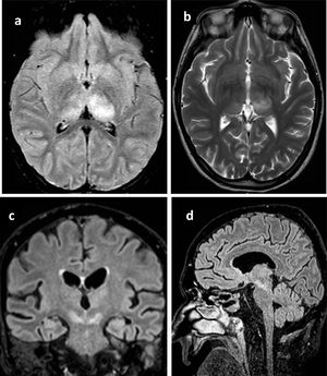
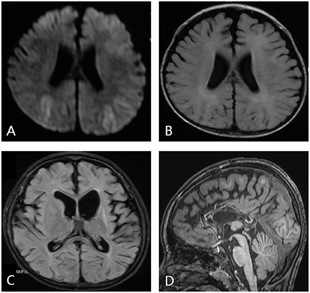
(a) Axial trace diffusion-weighted image obtained 24 h after the onset ...
Axial FLAIR ( A ) and axial T2 fast spin-echo ( B ) images show lesions ...
Infections of the Developing and Mature Nervous System | Radiology Key
The cumulative incidences of all CNS complications and PRES at days 30 ...
West Nile virus encephalitis outbreak in the Guadalquivir river basin ...
MRIs of a 10-month-old girl (patient 4) who was left with severe ...
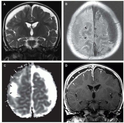
Representative imaging findings from 4 cases of PRES: (a) Axial T2 ...
Brain Imaging in Differential Diagnosis of Dementia - Practical Neurology
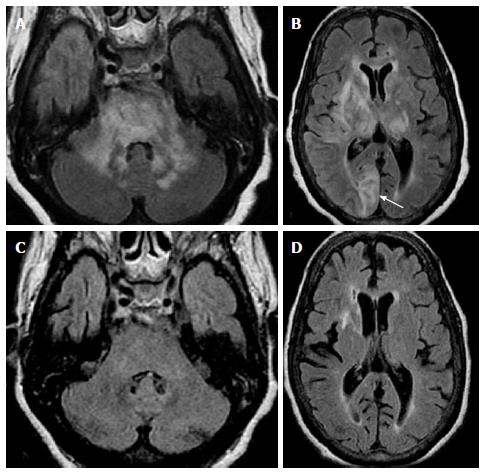
A) Axial T2-weighted image shows bilateral hyperintense signal changes ...
Clinical and genetic analysis of two Chinese infants with Mabry ...
Axial (A,B) and coronal (C,D) MRI of the brain/orbits prior to surgical ...
Cerebrovascular Disease | Radiology Key
Acute encephalitis with refractory, repetitive partial seizures - Brain ...
Morphological and functional MRI, MRS, perfusion and diffusion changes ...
The severity score of enlarged perivascular spaces in basal ganglia. a ...
Cerebrotendinous Xanthomatosis ataxia responsive to CDCA and Riluzole ...
Indications and Imaging Modality of Choice in Pediatric Headache ...
Restricted Diffusion in Vanishing White Matter | JAMA Neurology | JAMA ...
HHV-7–related encephalitis in 2-year-old girl with febrile convulsions ...
Axial T2-weighted MRI of the brain in July of 2014 showing hyperintense ...
1 MRI of case 1: FLAIR visible hippocampal, thalamic, and midbrain high ...
Infantile-onset Alexander disease in a child with long-term follow-up ...
Brain MRI performed 2 months after treatment with steroid. Multiple ...
(PDF) Three-dimensional brain MRI for DBS patients within ultra-low ...
Melanoma brain metastases were detected principally on T2*-weighted ...
Brain MRI displaying multifocal hyperintense signal changes in ...
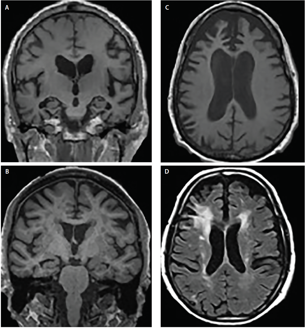
| (A) Magnetic resonance brain images with fluid attenuated inversion ...
Very late-onset mitochondrial cytopathy featuring epilepsia partialis ...
Frontiers | Pediatric epilepsy surgery in patients with Lennox-Gastaut ...
a and b: Axial, FLAIR MRI image of a NBD patient presenting ...
Propionic Acidemia: Case Report and Review of Neurologic Sequelae ...
A , Axial T2-weighted image shows a right frontotem- poral lesion with ...
Book DRPL (Dentatorubral-pallidoluysian Atrophy) Analysis | Test Price ...
Acute toxic encephalopathy due to the ingestion of Rhus extract ...
Middle cerebellar peduncles: Magnetic resonance imaging and ...
Cerebral-Infarction-ICD-10
Thalamic-Infarction
Basal-Ganglia-Infarction-ICD-10
Right-Thalamic-Stroke-ICD-10
Ischemic-Infarction-ICD-10
ICD-10-Infarction-R-Thalamus
Cerebellar-Infarction-ICD-10
Left-Thalamic-Infarction
Acute-Thalamic-Infarction
Posterolateral-Thalamic-Infarction
Thalamic-Infarction-ICD-10-Code
Acute-Thalamic-Infarction-Gross-Photo
Posterior-Circulation-Infarction-ICD-10
Right-PCA-Distribution-Infarction-ICD-10
Thalamic-Hemorrhage-ICD-10
Thalamic-Infarction-MRI-Diffusion


%20Analysis.webp)