Please enter url.
Login
Logout
Please enter url.
Radiographic images of brain disorders associated with Van Der Knaap ...
researchgate.net
source
Comments
Understanding the association of neurocysticercosis and mesial temporal ...
MR images obtained on day 5. AD, On axial T2WI, the lesions appear ...
A 41-year-old woman with barotrauma of the bilateral middle ears and ...
Glutaric aciduria type 1. A 7-month child with macrocrania and ...
Cerebral MRI abnormalities associated with vigabatrin therapy - Pearl ...
Brain MRI shows T2 hyperintensity ( arrows ) on the dorsal midbrain ( a ...
Claude's Syndrome Associated with Neurocysticercosis – ScienceOpen
HHV-7–related encephalitis in 2-year-old girl with febrile convulsions ...
Brain Imaging in Differential Diagnosis of Dementia - Practical Neurology
(a) Sagittal CT image in an 11-year-old male shows a pineal mass that ...
Infections of the Developing and Mature Nervous System | Radiology Key
Neuroimaging in MIRAS disease (POLG mutation) and PENDRED syndrome ...
Clinical variables associated with Rasmussen encephalitis and cortical ...
MRIs of a 10-month-old girl (patient 4) who was left with severe ...
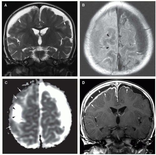
| Brain MRI. Axial T2 (A-C) and diffusion (D-F) showing swelling and ...
Deep Medullary Vein Involvement in Neonates with Brain Damage: An MR ...
Patient 1. Images at presentation (A) and follow-up 5 (B-E) and 45 (F ...
Bilateral basal ganglia lesions at presentation (a–e): hypodense on CT ...
(PDF) Bilateral Thalamic Infarction and DSA Demonstrated AOP after ...
Recent history of varicella-zoster virus infection in a 4-year-old boy ...
T1-weighted image obtained 2 months after exposure to carbon monoxide ...
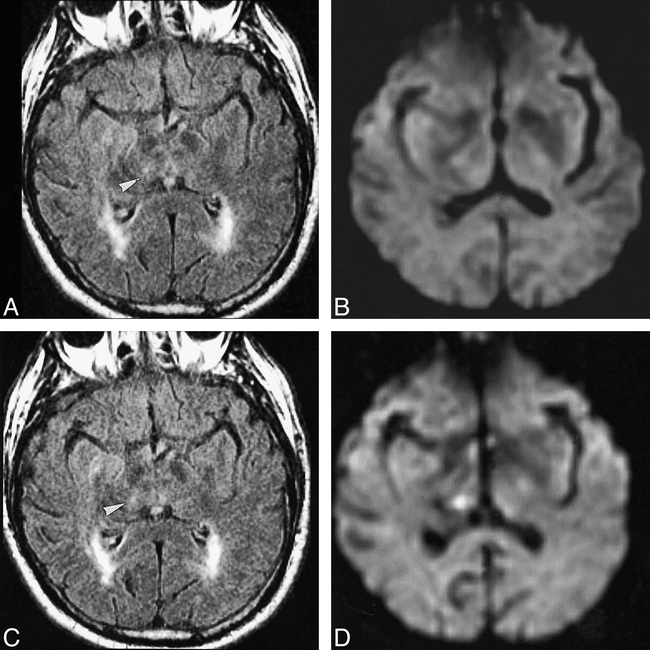
MR imaging of patient 1 on day 4 ( A and B ), day 6 ( C Ϫ E ), and day ...
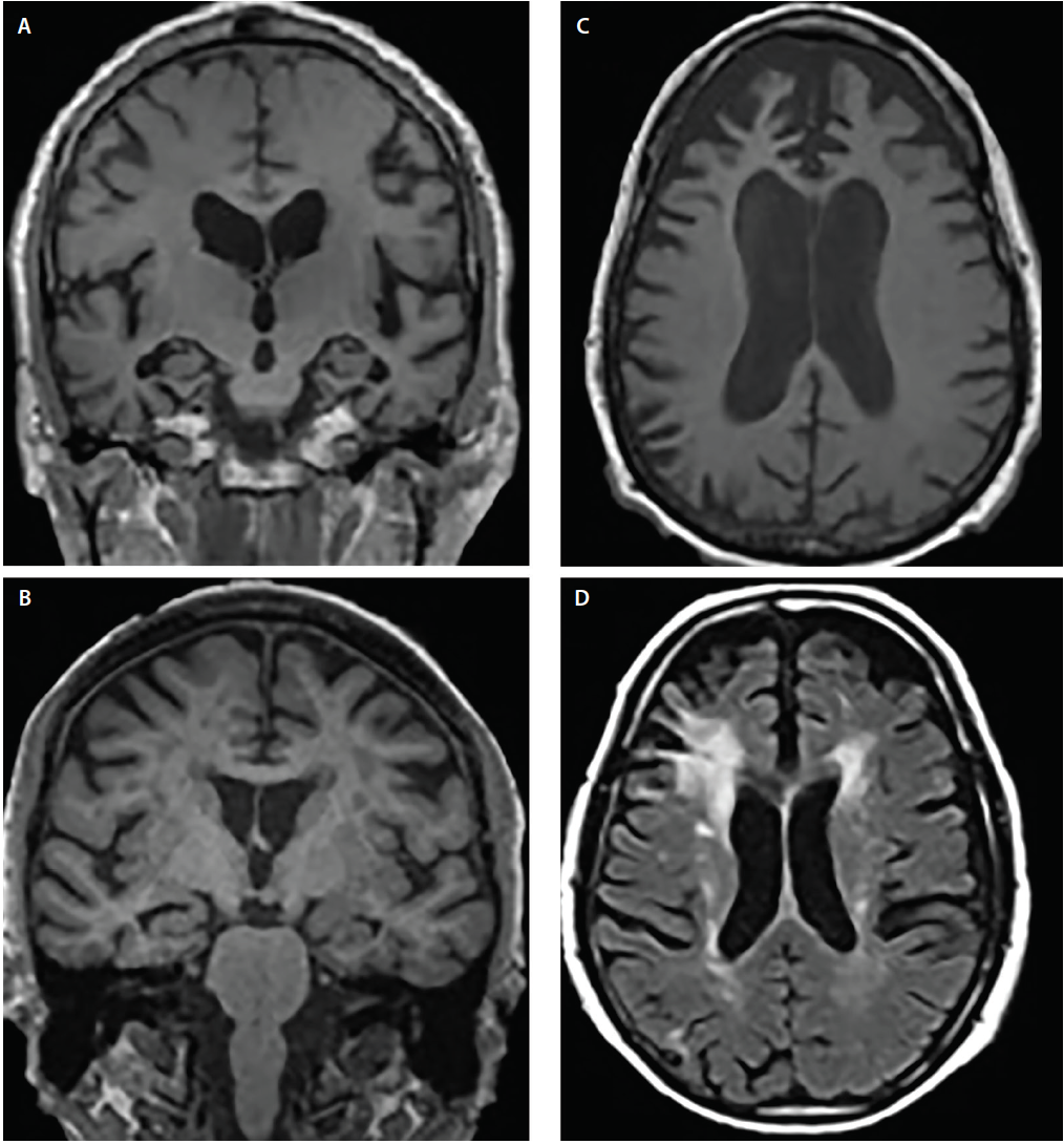
(a and b) Follow-up brain MRI after 2 months, partial resolution of ...
High-resolution, T2-weighted, FSE MRI (voxels of 0.47 mm ϫ 0.47 mm ϫ 2 ...
Axial T2-weighted MRI of the brain in July of 2014 showing hyperintense ...
(PDF) IS-029 Neonatal Stroke
Axial FLAIR (A) and diffusionweighted images (B) from the initial MR ...
Acute encephalitis with refractory, repetitive partial seizures - Brain ...
Figure2.Magnetic resonance imaging of cerebral diffusion-weighted ...
Brain imaging. Axial T2 FLAIR images (A and B), and coronal T2 images ...
[PDF] Middle interhemispheric variant of holoprosencephaly | Semantic ...
MRI brain. (A) Diffusion weighted image-revealing restriction of ...
Infantile-onset Alexander disease in a child with long-term follow-up ...
False-negative Diffusion-weighted MR Findings in Acute Ischemic Stroke ...
Human-Brain-Chart
Human-Brain-Mind
Busy-Brain
Dr-Space-Brain
Human-Brain-Anatomy
Brain-Stem-Model
Brain-Anatomy-Cartoon
Brain-Stem-Diagram
Brain-Gym-Exercises
Brain-Anatomy-and-Physiology
Brain-Amygdala-Prefrontal-Cortex
Dr-Brain-and-Brain-2
Frontal-Lobe-Brain-Anatomy
Brain-Anatomy-and-Functions
Amygdala-and-Hippo-Campus
Simple-Brain-Diagram


![[PDF] Middle interhemispheric variant of holoprosencephaly | Semantic ...](https://d3i71xaburhd42.cloudfront.net/a3849b2dbf40041d9030f26a86d2769f639fc64c/2-Figure2-1.png)