Please enter url.
Login
Logout
Please enter url.
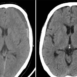
CT scans of the brain at SLE diagnosis (A) and current presentation (B ...
researchgate.net
source
Comments
CT scan brain plain showing bilateral thalamic hemorrhages | Download ...
(a) Initial CT scan showing left CSDH and (b) CT scan after irrigation ...
CT performed on day 5 shows low attenuation areas in subcortical white ...
(PDF) Neuroimaging Studies in Carbon Monoxide Intoxication
Right-hemisphere periventricular rosary-like pattern calcified lesions ...
Luria Nebraska Neuropsychological Battery-Clinical Scales | Download ...
Brain CT on admission showed an IVH in both lateral ventricles with ...
(PDF) Benign external hydrocephalus
Descending transtentorial herniation. A right temporal large traumatic ...
(a and b) Computed tomography brain showing a chronic subdural hematoma ...
Plain X rays of skull. (a) and (b) Curvilinear midline calcification in ...
Brain CT on admission showed an IVH in both lateral ventricles with ...
SIHE: After falling from standing position, CT images within 3 hours ...
Axial sections of brain MRI. Fluid attenuation inversion recovery ...
Radiologically suspected low grade gliomas (WHO I° & II°) | Download Table
-TC crânio-encefálica (2010) revelando calcificação bilateral dos ...
Cortical gyral enhancement in embolic cerebral infarction in a ...
Cortical venous thrombosis. A, Axial GRE T2 image shows right central ...
(PDF) Primary osteosarcoma of frontal bone: A case report and review of ...
CT scan performed 1 month after the SAH with a chronic subdural ...
A non-contrast computed tomography scan demonstrates acute large ...
Traumatic intracerebral hematoma. Axial non-enhanced CT scan shows a ...
Representative images of SAH with a CT scan. SAH, subarachnoid ...
(PDF) Supratentorial subdural hematoma following microvascular ...
Illustration of Case 1, progression of intracranial hemorrhage in 2 ...
Radiological Anatomy: Parieto-Occipital Sulcus - Stepwards
(PDF) Transient Obstructive Hydrocephalus due to Intraventricular ...
Brain MRI, the T2W image. There is no abnormal signal of the brain ...
(PDF) Endoscopy in the treatment of slit ventricle syndrome
Images on cranial CT: 1a (Right). Lacunar infarct in the left thalamus ...
CT scan in the axial plane showing extensive bilateral calcification of ...
A seemingly diffuse infiltrative non-enhancing mid brain lesion on the ...
Radiological Anatomy: Parieto-Occipital Sulcus - Stepwards
Computed tomography in axial plane shows bleeding into the subarachnoid ...
a and 2b: Non-contrast CT of the brain showing linear hyperdense areas ...
Axillary-Artery-and-Branches
Bronchial-Artery-CT
Thoracodorsal-Trunk
Axillary-Artery-Parts
Brachial-Artery-Angiogram
Pulmonary-Artery-CT
Subclavian-Axillary-Artery
Thoracic-Artery
Subscapular-Artery-CT
Lateral-Thoracic-Artery-CT
Popliteal-Artery-Aneurysm
Carotid-Artery-Anatomy-CT
Renal-Arteries-CT
Popliteal-Artery-Occlusion
Axillary-Artery-Cadaver
Thoracodorsal-Nerve-and-Artery