Please enter url.
Login
Logout
Please enter url.
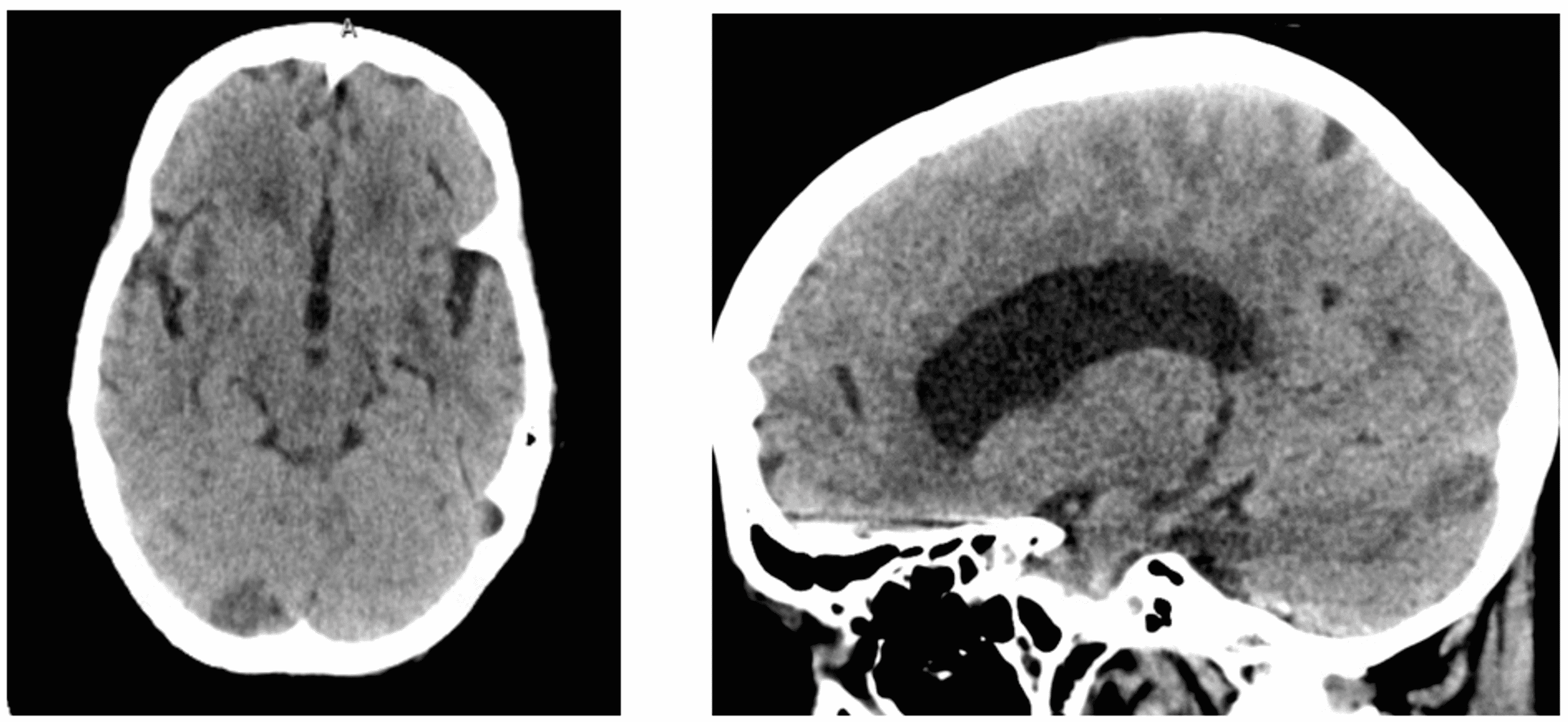
SIHE: After falling from standing position, CT images within 3 hours ...
researchgate.net
source
Comments
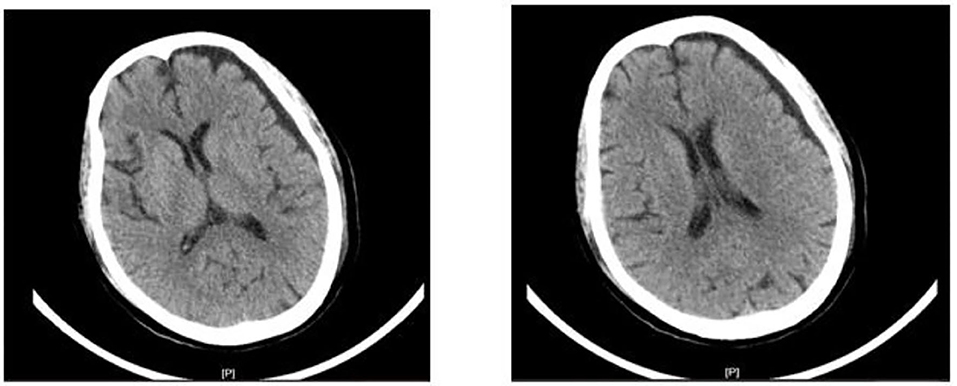
(a) Initial CT scan showing left CSDH and (b) CT scan after irrigation ...
Axial Brain CT scan of the patient Suspicious hypo density focus in ...
(PDF) Primary osteosarcoma of frontal bone: A case report and review of ...
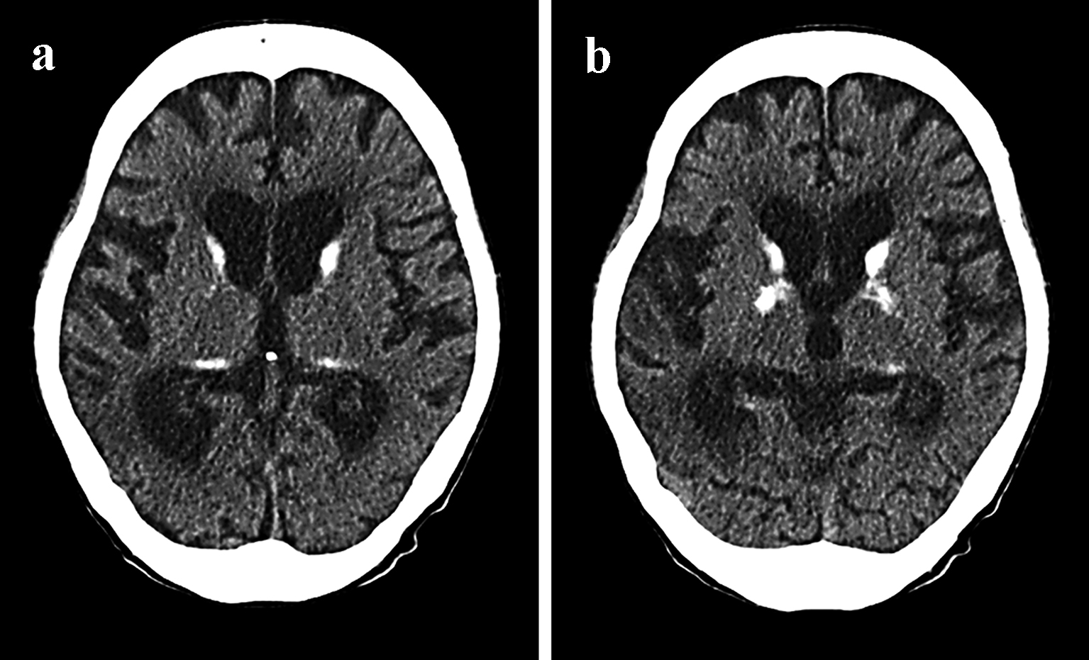
a . Bilateral basal ganglia calcification. b . Bilateral basal ganglia ...
Axial CT scans. a A 35-year-old male with the choroid plexus of the ...
MSCT of the brain. Measurement for calculating the index of the ...
CT scan showed a small left frontal subcortical hematoma (Fig 2, A ...
(PDF) The risk of ventricular catheter misplacement and intracerebral ...
Unilateral brain oedema related to focal status epilepticus | BMJ Case ...
Axial CT images at the level of the middle cerebellar peduncles show ...
MRI of brain enhanced with contrast agent showing ventricular ...
(A) Region of interest on the torcular Herophili; the patient is ...
Brain CT on admission showed an IVH in both lateral ventricles with ...
CT scan of the head. There is a mass effect on the right portion of the ...
Axial (A) and sagittal (B) CT angiograms show a giant arachnoid ...
Cureus | Cryptococcus Infection in an Immunocompetent Patient
CT scans of the brain at SLE diagnosis (A) and current presentation (B ...
On the day following endovascular treatment, computed tomography (CT ...
SIHE: After falling from standing position, CT images within 3 hours ...
Frontiers | Case report: Blood purification effectively relieves ...
Diagnostics | Free Full-Text | A Rare Case of Aphasia Caused by Delayed ...
Traumatic intracerebral hematoma. Axial non-enhanced CT scan shows a ...
A non-contrast computed tomography scan demonstrates acute large ...
A, Nonenhanced cranial CT showing right‐sided MCA territory infarct. B ...
Importance of Distinguishing Between Mitochondrial Encephalomyopathy ...
(PDF) Usefulness of embolization of the middle meningeal artery for ...
Pre- ( A ) and postcontrast ( B ) follow-up CT performed on day 24 ...
The modified Rankin Scale (mRS) | Download Scientific Diagram
Right-hemisphere periventricular rosary-like pattern calcified lesions ...
CT-scan of a number of control individuals. | Download Scientific Diagram
Serie ósea: esclerosis de huesos tubulares (a, b) con fracturas de ...
Bilateral traumatic basal ganglia hemorrhage. - Abstract - Europe PMC
(PDF) Supratentorial subdural hematoma following microvascular ...
(PDF) Benign external hydrocephalus
Cortical venous thrombosis. A, Axial GRE T2 image shows right central ...