Please enter url.
Login
Logout
Please enter url.
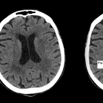
Images on cranial CT: 1a (Right). Lacunar infarct in the left thalamus ...
researchgate.net
source
Comments
(PDF) Traumatic surdural hygroma - Five cases with changed density and ...
Post-operative computed tomography scan of patient demonstrating small ...
(PDF) Benign external hydrocephalus
Radiological Anatomy: Parieto-Occipital Sulcus - Stepwards
Axial sections of brain MRI. Fluid attenuation inversion recovery ...
Radiological Anatomy: Sylvian Fissure - Stepwards
Brain CT on admission showed an IVH in both lateral ventricles with ...
(PDF) Neuroimaging Studies in Carbon Monoxide Intoxication
CT performed on day 5 shows low attenuation areas in subcortical white ...
CT scan brain plain showing bilateral thalamic hemorrhages | Download ...
CT scans of the brain at SLE diagnosis (A) and current presentation (B ...
Chronic calcified subdural hematoma of the frontoparietal right ...
(PDF) Contrast-induced encephalopathy after coronary angioplasty and ...
During follow-up, MRI scans showed a porencephalic cyst at the site ...
(a) Nonenhanced computed tomography brain showing thrombosed cortical ...
-Enhanced CT of tubercular meningitis with perivascular inflammation ...
Illustration of Case 1, progression of intracranial hemorrhage in 2 ...
Two transverse computed tomography slices on the frontal lobe and near ...
Radiological Anatomy: Parieto-Occipital Sulcus - Stepwards
Computed tomography in axial plane shows bleeding into the subarachnoid ...
(A) Magnetic resonance axial section T2* GRE sequence with extensive ...
Head CT showed slightly lower parenchyma density in both cerebral ...
Acute cerebral infarction is seen in the right fronto-temporal lobe ...
1: The central nervous system (CNS) and peripheral nervous system ...
Subependymal heterotopia. CT scan shows multiple heterotopic gray ...
Viewing playlist: Traumatic intracranial haemorrhage | Radiopaedia.org
(a) Preoperative computed tomography reveals right thalamic hemorrhage ...
| Post-procedure computed tomography scans showing different severity ...
CT scan performed 1 month after the SAH with a chronic subdural ...
EPOS™ - C-2725
Left Sylvian artery stroke. (A) Non-contrast CT weighted sum image ...
Shree DINESH | Director of Spine Surgery | FRCS(Surgical Neurology ...
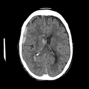
48 years old man with increased intra cranial pressure. Axial cranial ...
A. Computed tomography scan showing a mass at the cardia, which was ...
CT scan (December 18, 2018), with no contrast, showing a modest ...