Please enter url.
Login
Logout
Please enter url.
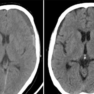
CT scan brain plain showing bilateral thalamic hemorrhages | Download ...
researchgate.net
source
Comments
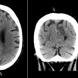
CT scan sagittal reconstruction showing extensive thalamic hemorrhages ...
Optimal window settings. (a) Standard window setting for soft tissue or ...
CT scans of the brain at SLE diagnosis (A) and current presentation (B ...
Expansile pontine lesion typical of a diffuse brain stem glioma on (a ...
Descending transtentorial herniation. A right temporal large traumatic ...
CT performed on day 5 shows low attenuation areas in subcortical white ...
(PDF) Benign external hydrocephalus
Right-hemisphere periventricular rosary-like pattern calcified lesions ...
(a) Initial CT scan showing left CSDH and (b) CT scan after irrigation ...
A, Nonenhanced cranial CT showing right‐sided MCA territory infarct. B ...
(a) Preoperative computed tomography reveals right thalamic hemorrhage ...
A non-contrast computed tomography scan demonstrates acute large ...
Non-contrast enhanced CT scan at the level of roof of lateral ventricle ...
Patient with a ruptured anterior communicating artery aneurysm and ...
(a and b) Computed tomography brain showing a chronic subdural hematoma ...
Axial CT scans. a A 35-year-old male with the choroid plexus of the ...
Cortical gyral enhancement in embolic cerebral infarction in a ...
(PDF) Isolated Adrenocorticotropic Hormone Deficiency Following Chronic ...
CT scans of Case 6. Acute subdural hematoma was evacuated by ...
Initial axial non contrast CT scan showing an acute left sided subdural ...
Axial CT images at the level of the middle cerebellar peduncles show ...
(PDF) Primary osteosarcoma of frontal bone: A case report and review of ...
(PDF) Neuroimaging Studies in Carbon Monoxide Intoxication
(PDF) Temporal fossa arachnoid cyst presenting with bilateral subdural ...
(PDF) Congenital and perinatal cytomegalovirus infection
Cortical venous thrombosis. A, Axial GRE T2 image shows right central ...
-TC crânio-encefálica (2010) revelando calcificação bilateral dos ...
A 39-year-old man with hyperacute subarachnoid hemorrhage caused by a ...
Initial brain computed tomography (CT) showed a 10.0 × 5.3 × 8.0 cm ...
Cranial CT scan showed hypodense lesions in the left frontal lobe and ...
abnormal mri brain Herpes encephalitis - Radiology Imaging
Radiology MRI
CT scan on hospital admission (a) demonstrating herniation of cerebral ...
(PDF) Assessment and treatment of childhood topographical ...
Representative images of SAH with a CT scan. SAH, subarachnoid ...
Punctate-Hemorrhage
Thalamic-Infarct
Left-Thalamic-Infarct
Right-Thalamic-Stroke
Putaminal-Hemorrhage
Parenchymal-Hemorrhage
Hypertensive-Hemorrhage
Thalamic-Stroke-Syndromes
Basal-Ganglia-Hemorrhage
Thalamic-Aphasia
Thalamic-Ich
Thalamic-Vascular-Supply
Thalamic-Lesion
Thalamic-Stroke-MRI
Intracranial-Brain-Hemorrhage
Bilateral-Thalamic-Stroke