Please enter url.
Login
Logout
Please enter url.
Disorders of Brain Development | Radiology Key
radiologykey.com
source
Comments
Subcortical Ischemic Vascular Dementia - Neurologic Clinics
(a) T1-weighted axial MRI brain, reveals right cerebral hemispheric ...
Subcortical Low Intensity on MR Images of Meningitis, Viral ...
fig1:Assessment of brain tissue injury after moderate hypothermia in ...
Case 1-2022: A 67-Year-Old Man with Motor Neuron Disease and Odd ...
Examples of the three ordinal categories of enlarged Virchow-Robin ...
Transverse T1-weighted magnetic resonance images of the brain. Notes ...
Explore the Intricacies of Cortical Highlighting
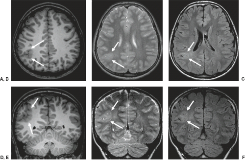
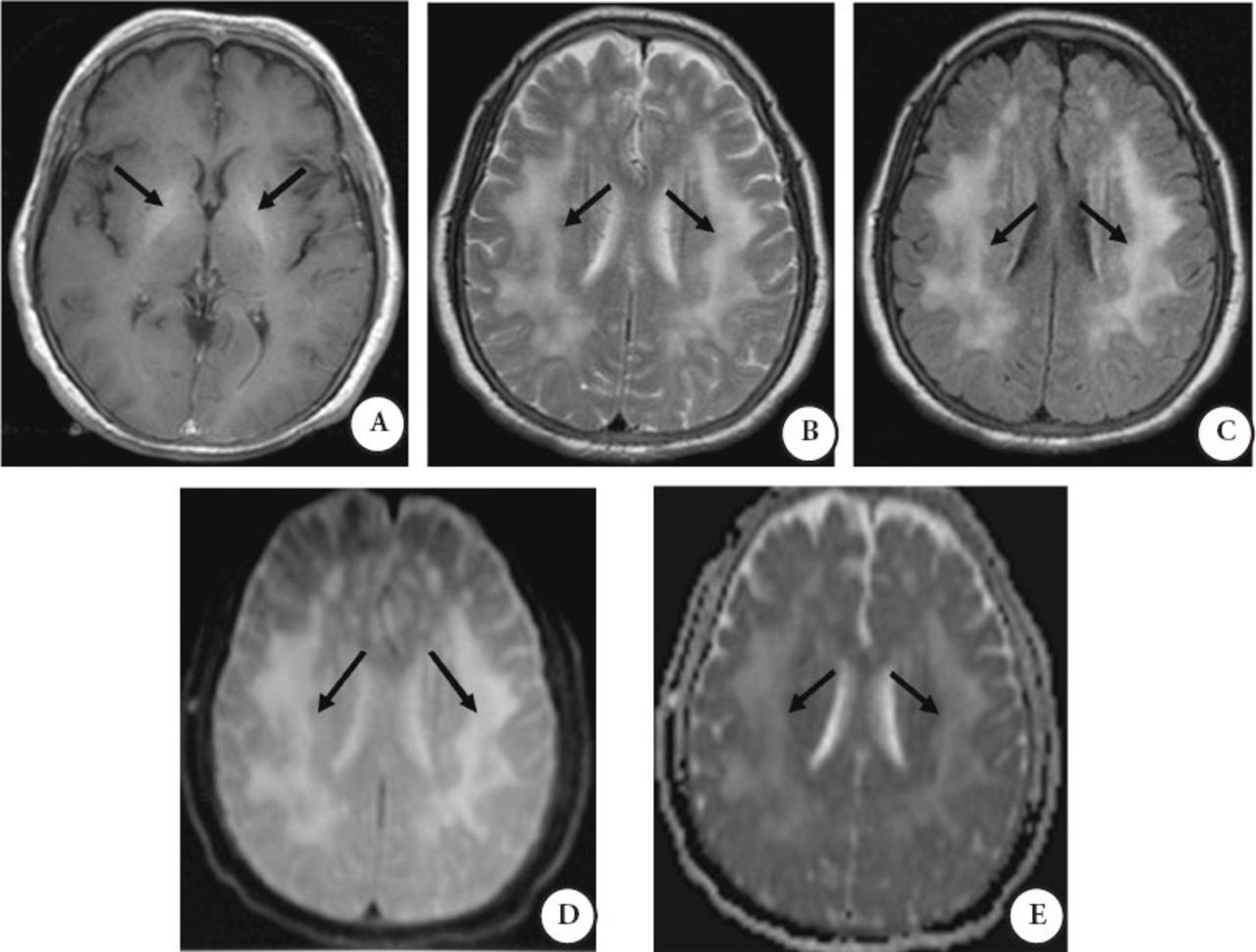
Brain MRI of two patients (Patient 1; A–C; Patient 2; D, E) with ...
Shielded Gradients. And The General Solution To The Near Field Problem ...
Central nervous system anomalies on MRI T2-weighed images in two ...
(PDF) Cerebral angiitis in four patients with chronic GVHD
Hepatic encephalopathy coexists with acquired chronic hepatocerebral ...
Posterior Reversible Encephalopathy Syndrome: Utility of Fluid ...
Malik GHANNAM | Vascular Neurology Fellow | MBBCh | University of Iowa ...
Subtraction MRI in a patient with relapsing–remitting multiple ...
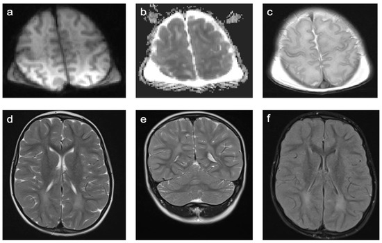
Children | Free Full-Text | Characterization of MRI White Matter Signal ...
Brain MRI showing high signal intensity lesions on both FLAIR (upper ...
Brain MRI made at the age of 13 months (images A, B, and C) and at the ...
Iron Oxide Particle-Enhanced MRI Suggests Variability of Brain ...
-Axial MRI images, Diffusion weighted images (DWI) long b value (1000 ...
Silent Brain Infarcts | Stroke
Illustration of lesion segmentation. (A) GM lesion segmentation, (B ...
Cerebral Amyloid Angiopathy: Diagnosis, Clinical Implications, and ...
Figure 3 from The many faces of posterior reversible encephalopathy ...
Magnetic resonance imaging of the brain revealing cortical atrophy that ...
Blood-brain barrier (BBB) breakdown and neurological complications. (A ...
Examples of the three ordinal categories of enlarged Virchow-Robin ...
2020–2021 BCSC Basic and Clinical Science Course™
| Brain magnetic resonance imaging (MRI) findings in patients 1 and 2 ...
Age-Related Total Gray Matter and White Matter Changes in Normal Adult ...
Parkinson’s Disease and Fabry Disease: Clinical, Biochemical and ...
Magnetic Resonance Image Brain Abnormalities in Myelin-Oligodendrocyte ...
Radiology Key
3D postcontrast FLAIR and postcontrast T1-weighted MR images in the ...
Sacroiliitis-Radiology
FCD-Brain
Cortical-Dysplasia-MRI
Fibrous-Cortical-Defect-MRI
Transmantle-Sign-Radiology
Focal-Cortical-Dysplasia-Radiology
Ffd-Dan-Radiology
Enchondroma-Femur-MRI
NonOssifying-Fibroma
Non-Ossifying-Fibroma-Femur
Mediastinal-Cyst-Radiology
Cortical-Stroke
Hippocampal-Sclerosis-MRI
Multiple-FCD-Radiology
Cortical-FCD-1-Radiology
Fibroma-No-Osificante