Please enter url.
Login
Logout
Please enter url.
(PDF) Cerebral angiitis in four patients with chronic GVHD
researchgate.net
source
Comments
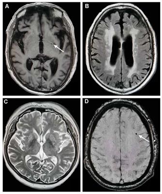
Magnetic resonance imaging (MRI) showing T2-weighted (a, d ...
Brain MRIs of the son before and after treatment of the... | Download ...
Subtraction MRI in a patient with relapsing–remitting multiple ...
MRI of Patient 2, revealing multiple lacunar infarctions of frontal ...
(A-H) Axial MRI after initial presentation: Diffuse periventricular and ...
Brain magnetic resonance imaging (MRI) of patients having MOG ...
Radiological progression by MRI of PML-associated brain changes from ...
| Brain MRI of the second and third attack. (A-C) Brain MRI ...
Head MRI on admission and day 21. (A-C) Head MRI on admission day. (A ...
| Example of MRI response and complication, in left temporal ...
Illustration of a disease assessment of progressive disease based on ...
| Brain magnetic resonance images of a 50-year-old woman with headache ...
Pedigree of the epilepsy family with Dyke-Davidoff-Masson syndrome. The ...
Summary of the RANO Response Criteria [14]. | Download Table
MRI of the brain, FLAIR sequence (TR/TE/FA: 7000/135/150). (A-C): Axial ...
| Brain MRI in a boy with CYP2U1 mutation. (A-C) Axial fluid-attenuated ...
Two exemplary MRI lesions after removal of ICP probe. Panel (A,E) show ...
Magnetic resonance imaging of the brain. Fluid-attenuated inversion ...
(PDF) Predominant Corticospinal Tract Involvement in a Late Infant with ...
(PDF) Current Opportunities for Clinical Monitoring of Axonal Pathology ...
Brain CT scan and MRI of a patient with LM intracranial listeria ...
Cranial MRI showing high intensity signal in in the splenium of the ...
Brain magnetic resonance imaging demonstrating characteristic findings ...
Example of deep grey matter T2 hypointensities of the caudate nuclei ...
Frontiers | Chorea-Acanthocytosis in a Chinese Family With a Pseudo ...
MRI images of brain lesion. MRI shows a well circumscribed cortical ...
1.5 T T2-weighted images with 5 mm slice thicknesses show basal ganglia ...
Frontiers | Total Cerebral Small Vessel Score Association With Hoehn ...
Magnetic resonance imaging results showing possible posterior ...
Magnetic resonance imaging of the brain revealing cortical atrophy that ...
Axial T1w +c craniocerebral MRI. (A) A baseline MRI showed a patch of ...
Development of CLN in Patient 3. (A) Fluid attenuation inversion ...
MRI appearance of unilateral right-sided polymicrogyria (PMG ...
Magnetic resonance images show bilateral atrophy of the head of the ...
Imaging of postoperative MRI. (a) Day 1 after surgery axial gadolinium ...


![Summary of the RANO Response Criteria [14]. | Download Table](https://www.researchgate.net/profile/Alexander-Radbruch/publication/51857202/figure/fig4/AS:277577425014789@1443191023458/T1-progress-without-previous-T2-progress-a-and-b-Recurrent-glioblastoma-after-initial_Q320.jpg)