Please enter url.
Login
Logout
Please enter url.
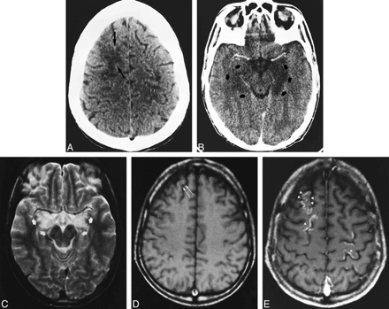
Magnetic Resonance Image Brain Abnormalities in Myelin-Oligodendrocyte ...
researchgate.net
source
Comments
JaypeeDigital | eBook Reader
Fluorescent Treponemal Antibody-Absorption Test | Semantic Scholar
The central nervous system | Radiology Key
Standardized brain MRI protocol to evaluate patients in whom multiple ...
Clinical presentation and pre-mortem diagnosis of variant Creutzfeldt ...
Intracranial Infection | Radiology Key
Posterior reversible encephalopathy syndrome: clinical and radiological ...
MRI findings in SANDO variety of the ataxia-neuropathy spectrum with a ...
Central Brain Herniation Secondary to JuvenileDiabetic Ketoacidosis ...
Everolimus for Subependymal Giant-Cell Astrocytomas in Tuberous ...
Children | Free Full-Text | Characterization of MRI White Matter Signal ...
MR Imaging of Cerebral Cortical Involvement in Aceruloplasminemia ...
Different types of PVWMH. Examples of frontal PVWMH with a smooth ...
Cortical Laminar Necrosis Caused by Immunosuppressive Therapy and ...
Figure 2 from Wilson's disease: An Indian perspective. | Semantic Scholar
Unenhanced head CT showing small hypodensities at the right caudate ...
Figure 1 from Alexia Without Agraphia as the Initial Manifestation of ...
Figure 2 from Gliomatosis cerebri mimicking acute viral encephalitis ...
1 MRI of case 1: FLAIR visible hippocampal, thalamic, and midbrain high ...
Cortical deafness of following bilateral temporal lobe stroke - Journal ...
MRA TOF sequence without contrast after the patient admission (A) shows ...
T2-weighted axial magnetic resonance imaging (MRI) scans (a, b) showing ...
Postoperative T2W axial (a-d) and T1W coronal (e-h) images reveal total ...
Genetic Analysis of Inherited Leukodystrophies: Genotype-Phenotype ...
A 5-year-old girl with left cerebellar JPA. Residual tumor failed to ...
Case 5. A: T 1 -weighted magnetic resonance (MR) image showing the ...
Figure 1 from Hemichorea-Hemiballism with a Diabetic Patient | Semantic ...
Brain MRI scan at 4 months of age with a typical pattern of AGS (A ...
Cerebral MRI, 2 months after onset of psychosis, epilepsy, and visual ...
| Brain magnetic resonance imaging (MRI) findings in patients 1 and 2 ...
Clinical presentation and imaging of general paresis due to ...
Imaging findings of intracranial calcifications. (A) Axial T1WI showed ...
Unusual Clinical and Magnetic Resonance Imaging Findings in a Family ...
Axial T1-weighted plain (a) and contrast (b) showing left... | Download ...
T2 weighted (a) and FLAIR (Fluid attenuated inversion recovery) MRI ...
Oligodendrocytes-Histology
Schwann-Cells
Astrocytes
Microglial-Cells
Myelin-Oligodendrocyte
Microglia
Satellite-Cells
Oligodendrocyte-Progenitor-Cells
Ependymal-Cells
Oligodendrocyte-Precursor-Cells
Neuroglia
Astrocytes-Function
Astrocytes-vs-Oligodendrocytes
Oligodendrocytes-Microscope
Oligodendroglioma
Glial-Cells