Please enter url.
Login
Logout
Please enter url.
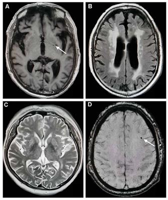
Subtraction MRI in a patient with relapsing–remitting multiple ...
researchgate.net
source
Comments
Standardized brain MRI protocol to evaluate patients in whom multiple ...
(A-H) Axial MRI after initial presentation: Diffuse periventricular and ...
CT and MRI brain of MELAS patients with nuclear gene mutations. Axial ...
Reoperation for Refractory Epilepsy in Childhood: A Second Chance for ...
Axial T1/SE magnetic resonance imaging (MRI) view of (a) a 25‐year‐old ...
Frontiers | Total Cerebral Small Vessel Score Association With Hoehn ...
Chronological changes of the brain MRI in case 3. At the age of ...
(PDF) Cerebral angiitis in four patients with chronic GVHD
| Sagittal STIR (A), sagittal T2 (B) and axial T2 (C) images of the ...
MRI appearance in a 45‑year‑old female patient with primary central ...
Figure 1 from Role of MRI in X-linked adrenoleukodystrophy—A case ...
MRI images showing acute haemorrhagic necrotising encephalopathy. (a ...
Magnetic resonance imaging of the brain revealing cortical atrophy that ...
Persistent falcine sinus and occipital sinus in case 2. (A, B) Sagittal ...
(A) T1, (B) T2, (C) diffusion weighted imaging, (D) susceptibility ...
Frontiers | Chorea-Acanthocytosis in a Chinese Family With a Pseudo ...
MRI findings in a male patient presented with glioblastoma multiforme ...
Brain magnetic resonance imaging (MRI). (A-F) Initial MRI evaluation ...
The sequences of the relevant areas of the ARFGEF2 gene are depicted ...
| Brain MRI of the second and third attack. (A-C) Brain MRI ...
Acute metronidazole-induced neurotoxicity: an update on MRI findings ...
MRI of Patient 2, revealing multiple lacunar infarctions of frontal ...
Brain MRI in AOA2 patients. Panels A-C show brain MRI images of patient ...
MRI of the brain of a patient with MLC (a-c) and MDC1A (d-f). The axial ...
Illustration of a disease assessment of progressive disease based on ...
Grading of degree of pulvinar hyperintensity in vCJD. Because only ...
| Results of head MRI. (A,B) Head MRI showed encephalatrophy in case 1 ...
Neuroradiologic imaging pattern of primary brain amyloidoma. The ...
Cranial MRI showing high intensity signal in in the splenium of the ...
(PDF) Expanding the spectrum of megalencephalic leukoencephalopathy ...
-Axial MRI images, Diffusion weighted images (DWI) long b value (1000 ...
Magnetic resonance imaging of the brain. Fluid-attenuated inversion ...
Figure 1 from A Case of Cerebral Granuloma and Optic Papillitis due to ...
Identifying relevant biomarkers of brain injury from structural MRI ...
a-d Right basal ganglia showing previous T2/FLAIR hyperintensity along ...
Cardiac-Amyloid-MRI
Cardiac-MRI-Amyloidosis
Cardiac-MRI-Indications
MRI-Protocols
MRI-Cardiac-Imaging
Cardiac-MRI-Sarcoidosis
Siemens-Cardiac-MRI
Cardiac-MRI-LGE
Cardiac-MRI-Procedure
Cardiac-Segments-MRI
Stress-Cardiac-MRI
Cardiac-MRI-Views
Cardiac-MRI-Machine
Normal-Cardiac-MRI
GE-MRI-Protocols
Brachial-Plexus-MRI-Protocol