Please enter url.
Login
Logout
Please enter url.
Perinatal Hypoxic Ischemic Encephalopathy
ar.inspiredpencil.com
source
Comments
fig1:Assessment of brain tissue injury after moderate hypothermia in ...
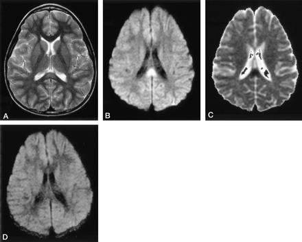
Brain MRI made at the age of 13 months (images A, B, and C) and at the ...
A 5-year-old girl with left cerebellar JPA. Residual tumor failed to ...
Patient 1: facial features with down-slanting palpebral fissures ...
Early Subacute Hematoma - Cytotoxic Edema - Mussen Healthcare
Neurosyphilis presenting with gummatous oculomotor nerve palsy ...
Isolated Cortical Visual Loss With Subtle Brain MRI Abnormal ...
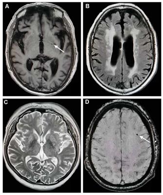
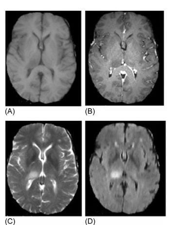
(A) T1* gradient echo MRI. Images show abnormal low signal bilateral ...
10 Prominent Perivascular Space | Radiology Key
High-resolution, T2-weighted, FSE MRI (voxels of 0.47 mm ϫ 0.47 mm ϫ 2 ...
Diagnosing Variant Creutzfeldt-Jakob Disease with the Pulvinar Sign: MR ...
Silent and apparent cerebral embolism after retrograde catheterisation ...
Vascular dementia - The Lancet
Intraventricular Hemorrhage - Cytotoxic Edema - Mussen Healthcare
Influenza-Associated Encephalitis/Encephalopathy with a Reversible ...
Bupropion Overdose Presenting as Status Epilepticus in an Infant ...
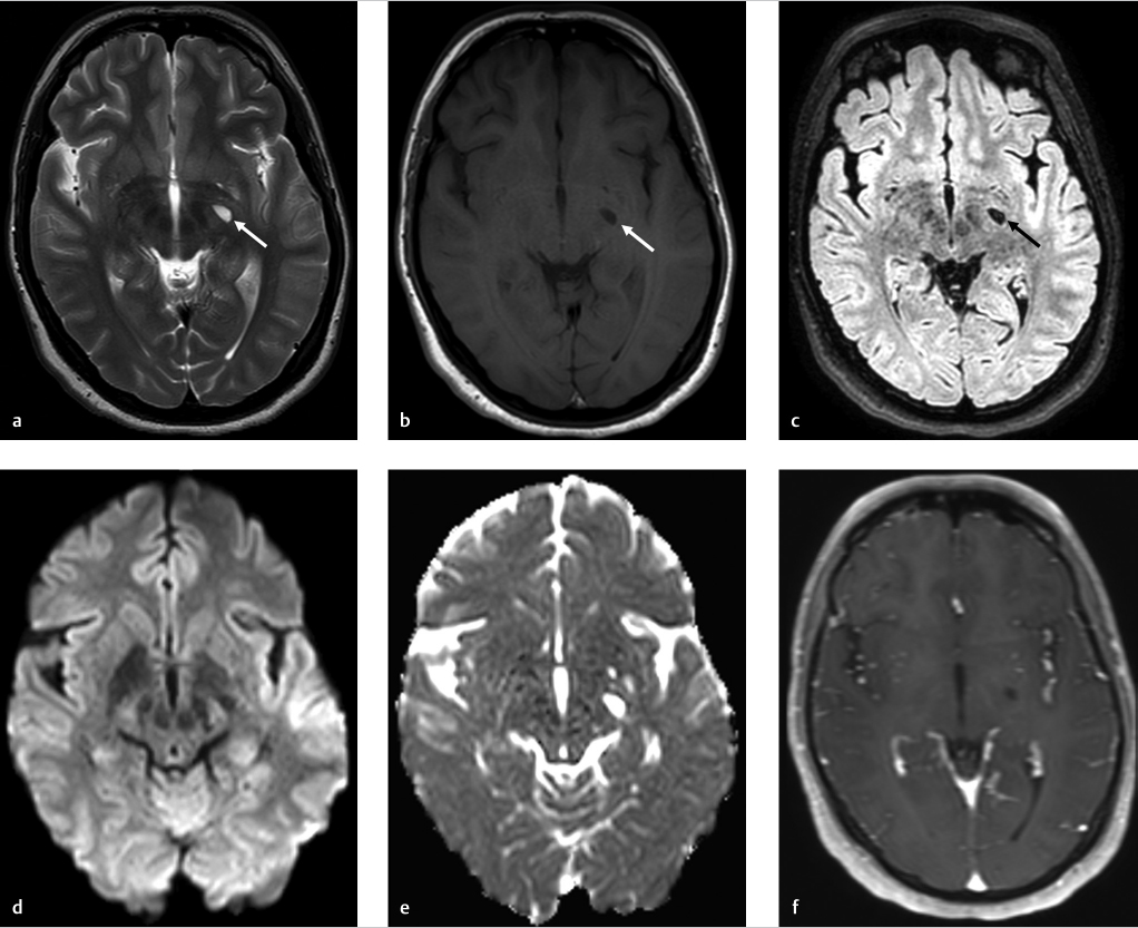
a focal restricted diffusion area adjacent to the lesion in the middle ...
Vigabatrin‐associated reversible MRI signal changes in patients with ...
Multiple ovoid multiple sclerosis lesions oriented perpendicular to the ...
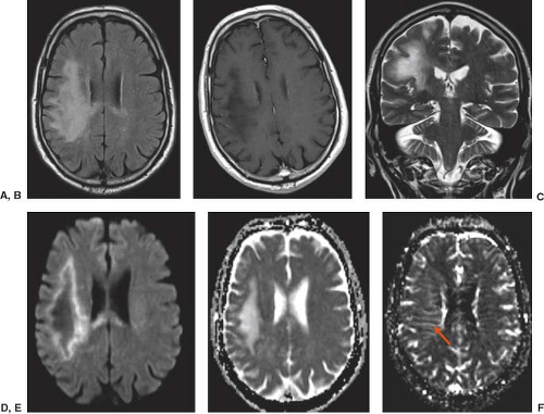
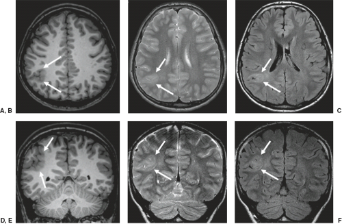
| Brain MRI. Axial T2 (A-C) and diffusion (D-F) showing swelling and ...
Childhood disorders of neurodegeneration with brain iron accumulation ...
Frontiers | Total Cerebral Small Vessel Score Association With Hoehn ...
Bilateral basal ganglia lesions at presentation (a–e): hypodense on CT ...
xmlinkhub
MRI in multiple sclerosis: current status and future prospects - The ...
Analysis of Cystic Intracranial Lesions Performed with Fluid-Attenuated ...
Headache, dizziness, sensorineural hearing loss, and bilateral leg ...
Lymphomatosis cerebri: a treatable cause of rapidly progressive ...
(a) Eleven-year-old male. T2W axial demonstrates numerous hypointense ...
MRI of Patient 2, revealing multiple lacunar infarctions of frontal ...
Intracranial Infection | Radiology Key
Disorders of Brain Development | Radiology Key
Case 38-2023: A 68-Year-Old Woman with Abnormal Movements and Confusion ...
Brain Tumour, MRI Images and CT Images | LaptrinhX
Coinfection of Japanese Encephalitis with Neurocysticercosis: An ...
Hypoxic-Injury
Hypoxic-Brain-Injury-CT
Anoxic-Brain-Injury-MRI
Hypoxic-Ischemic-Brain-Injury
Hypoxic-Cell-Injury
Hypoxic-Brain-Injury-at-Birth
Hypoxic-Brain-Damage
Brain-Hypoxia-Symptoms
Anoxic-Brain-Injury-CT-Scan
Severe-Anoxic-Brain-Injury
Anoxic-Brain-Injury-CT-Head
Cerebral-Hypoxia
Diffuse-Anoxic-Brain-Injury-On-MRI
Global-Brain-Hypoxia-MRI
Hypoxic-Ischemic-Encephalopathy
DWI-MRI-Hypoxic-Brain