Please enter url.
Login
Logout
Please enter url.
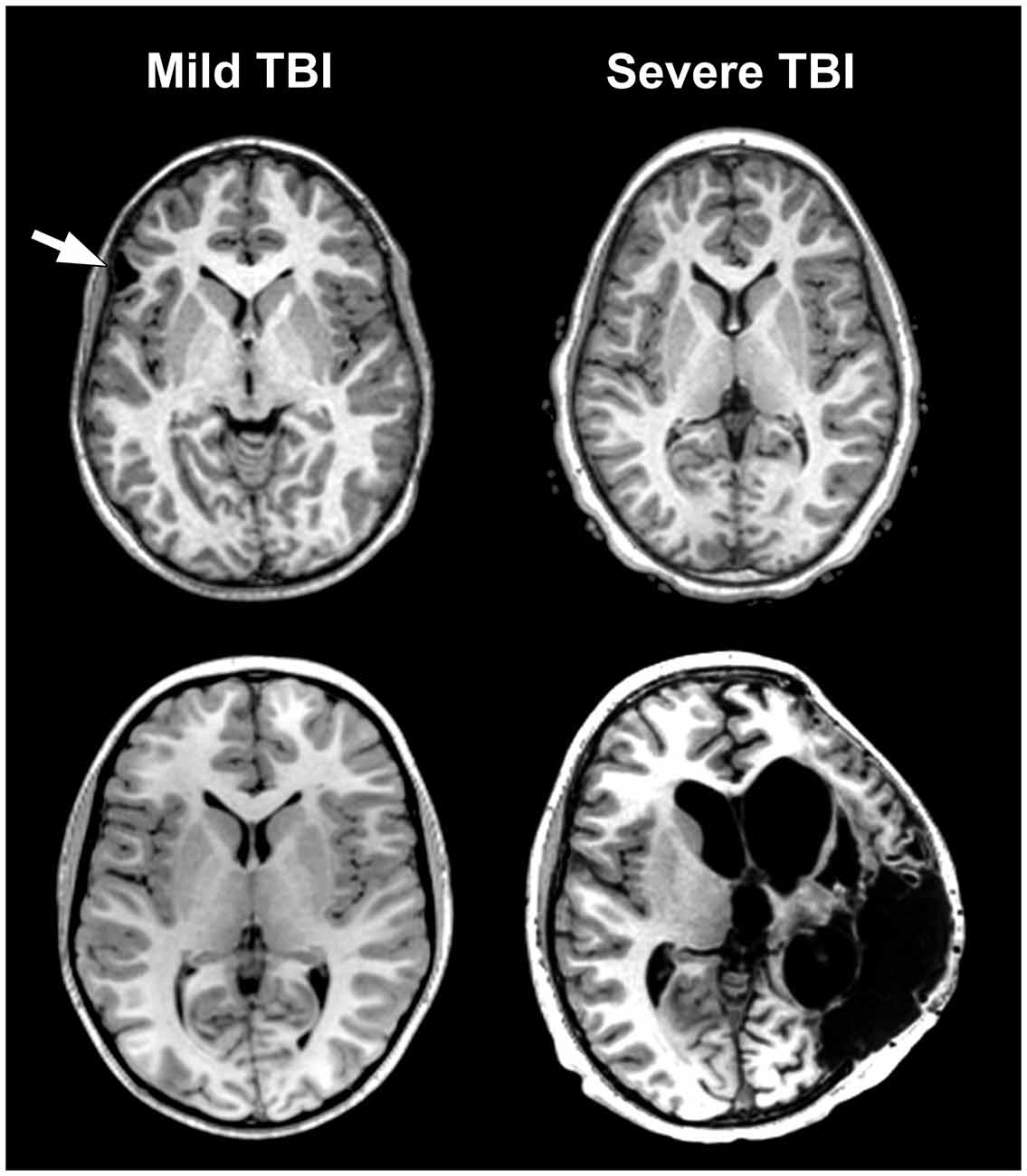
Traumatic Brain Injury Mri
ar.inspiredpencil.com
source
Comments
Frontiers | Systems Biology, Neuroimaging, Neuropsychology ...
Understanding White Matter Disease | Stroke
Diffuse Axonal Injury - Grading - Prognosis - TeachMeSurgery
Auditory processing disorder in patients with language-learning ...
Sagittal FSE (A) and high resolution axial (B) T2 weighted images ...
Examples of ADC measurements in the neonatal ADC map. This infant was ...
Frontiers | Fatal Outcome of European Tick-borne Encephalitis after ...
Diffusion MR Imaging of Hypoglycemic Encephalopathy | American Journal ...
Utility of diffusion-weighted imaging (DWI) and apparent diffusion ...
Figure 5 from Automatic curvilinear reformatting of three-dimensional ...
Imaging and CSF analyses effectively distinguish CJD from its mimics ...
Routine clinical brain MRI sequences for use at 3.0 Tesla - Lu - 2005 ...
Possible Creutzfeldt-Jakob Disease (Cjd): Case Report | Biomedres
Representatives images from brain MRI. Not the multiple lesions most ...
MRI-guided Focused Ultrasound Thalamotomy for Patients with Medically ...
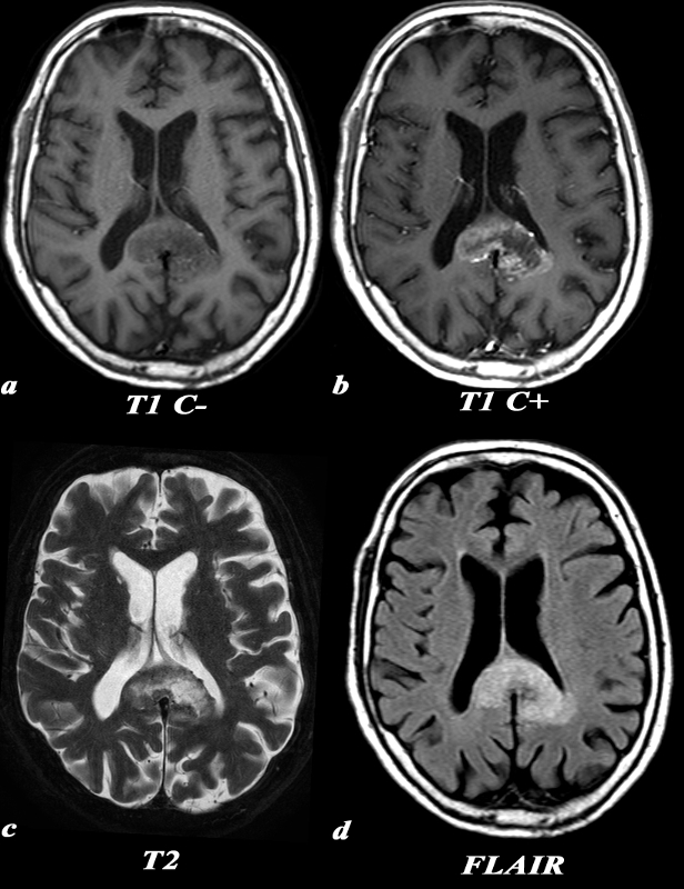
Corpus Callosum | Brain
Management of Epstein–Barr virus-related post-transplant ...
A 30‐Year Clinical and Magnetic Resonance Imaging Observational Study ...
Patient 3: Cavity, Edema, and Secondary Tumor Simulation, Fraction 1 ...
Cerebral Hyperperfusion Syndrome After Surgical Repair of Congenital ...
75 Best images about Multiple Sclerosis on Pinterest | Autoimmune ...
Brain Magnetic Susceptibility Changes in Patients with Natalizumab ...
Hyperglycemia and Hemorrhagic Transformation of Cerebral Infarcts | Stroke
| MRI of Patient 6, showing hyperintensity of the deep white matter on ...
Multiple Sclerosis Research: Rebranding MS as a Dementia
Posterior reversible encephalopathy syndrome (PRES) in a patient with ...
Leucoencephalopathy following abuse of sniffed heroin - Journal of ...
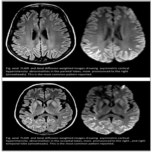
In images A – D the right of the brain is on the left side of the ...
Lissencephaly Mri
MR imaging of cerebral malaria in a child - European Journal of Radiology
Figure 54.2 from Brain CT and MRI: differential diagnosis of imaging ...
Physiology physics woven fine: Relaxation in the Nuclear Microcosm
Hereditary diffuse leukoencephalopathy with axonal spheroids caused by ...
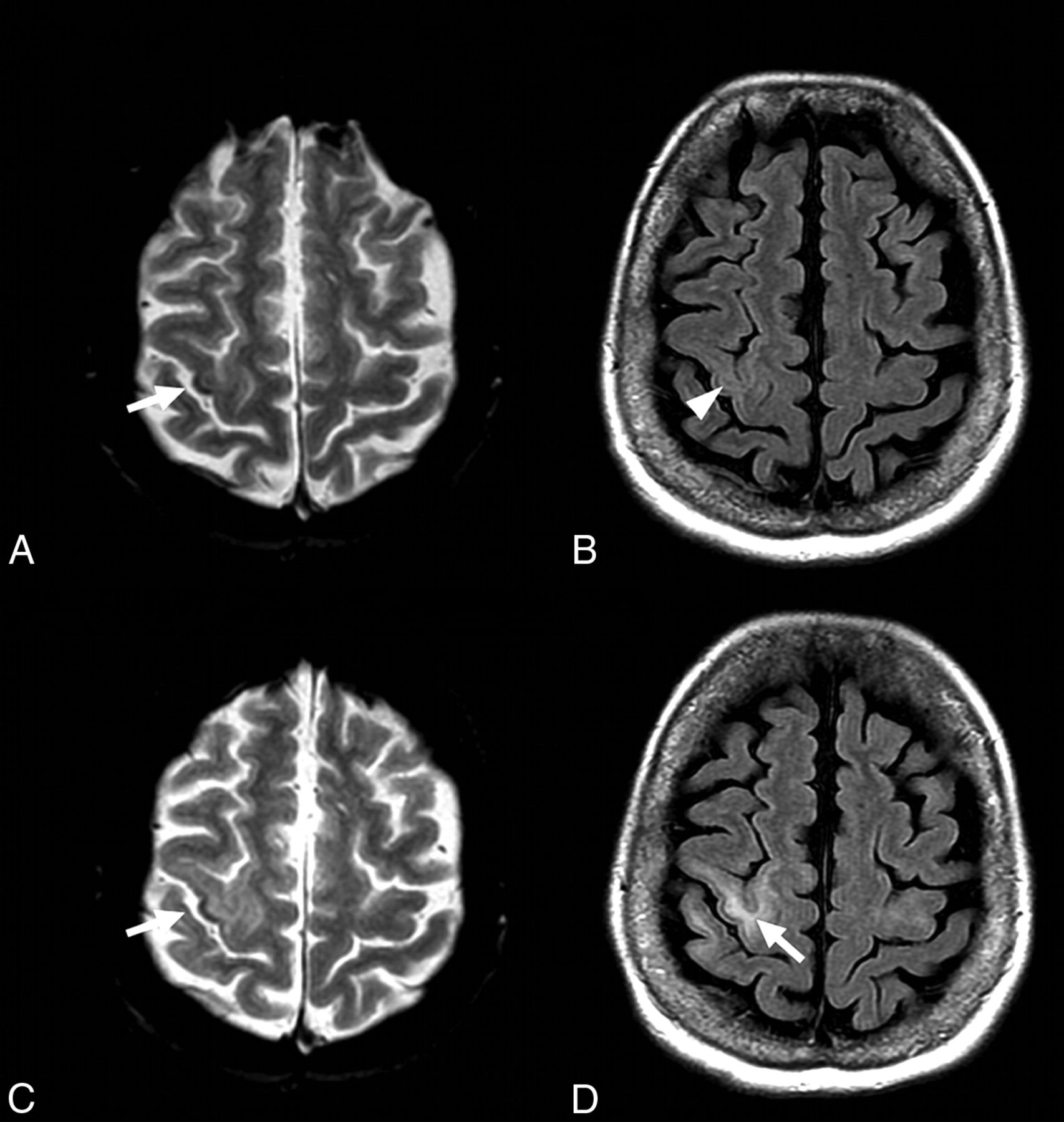
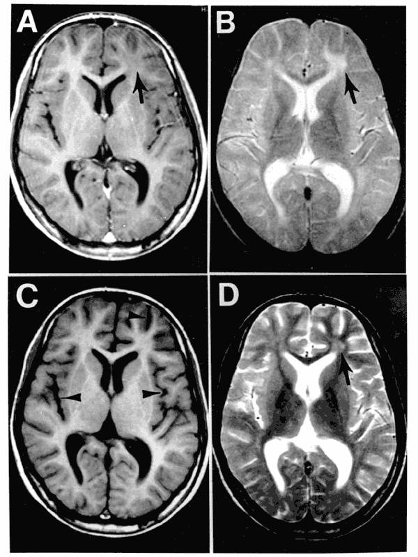
Axial T1- weighted (A, B) and T2- weighted (C, D) MR images in two ...
Lesions of seizure induced brain edema in a 64-year-old male. MRI and ...
Severity-Vs.-Priority
Asthma-Severity-Classification
Severity-Levels
Classifying-Asthma-Severity
Asthma-Severity-Criteria
COPD-Severity-Scale
Severity-Types
Hemophilia-Severity-Classification
Asthma-Severity-Classification-Chart
4-Classifications-of-Asthma
Pe-Severity-Classification
Asthma-Severity-Assessment
Asthma-Symptom-Severity
Asthma-Step-Chart
Disease-Severity
Bone-Loss-Severity-Classification