Please enter url.
Login
Logout
Please enter url.
Physiology physics woven fine: Relaxation in the Nuclear Microcosm
physiology-physics.blogspot.com
source
Comments
Figure 1 - Subacute Sclerosing Panencephalitis, a Measles Complication ...
Glutaric aciduria type 1. A 7-month child with macrocrania and ...
Cerebral toxoplasmosis responding to therapy. (A) Axial T2-weighted ...
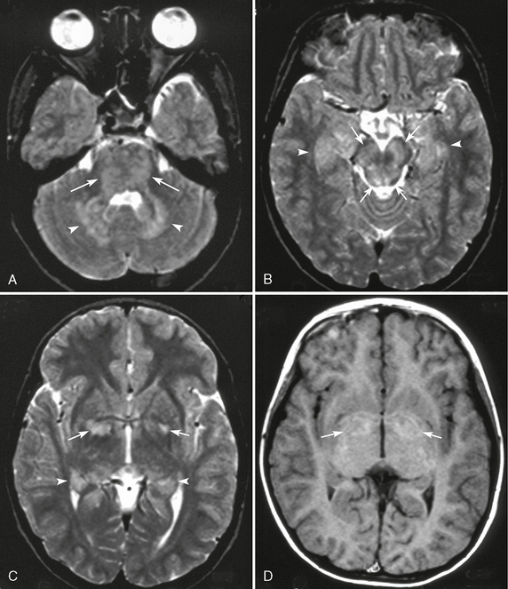
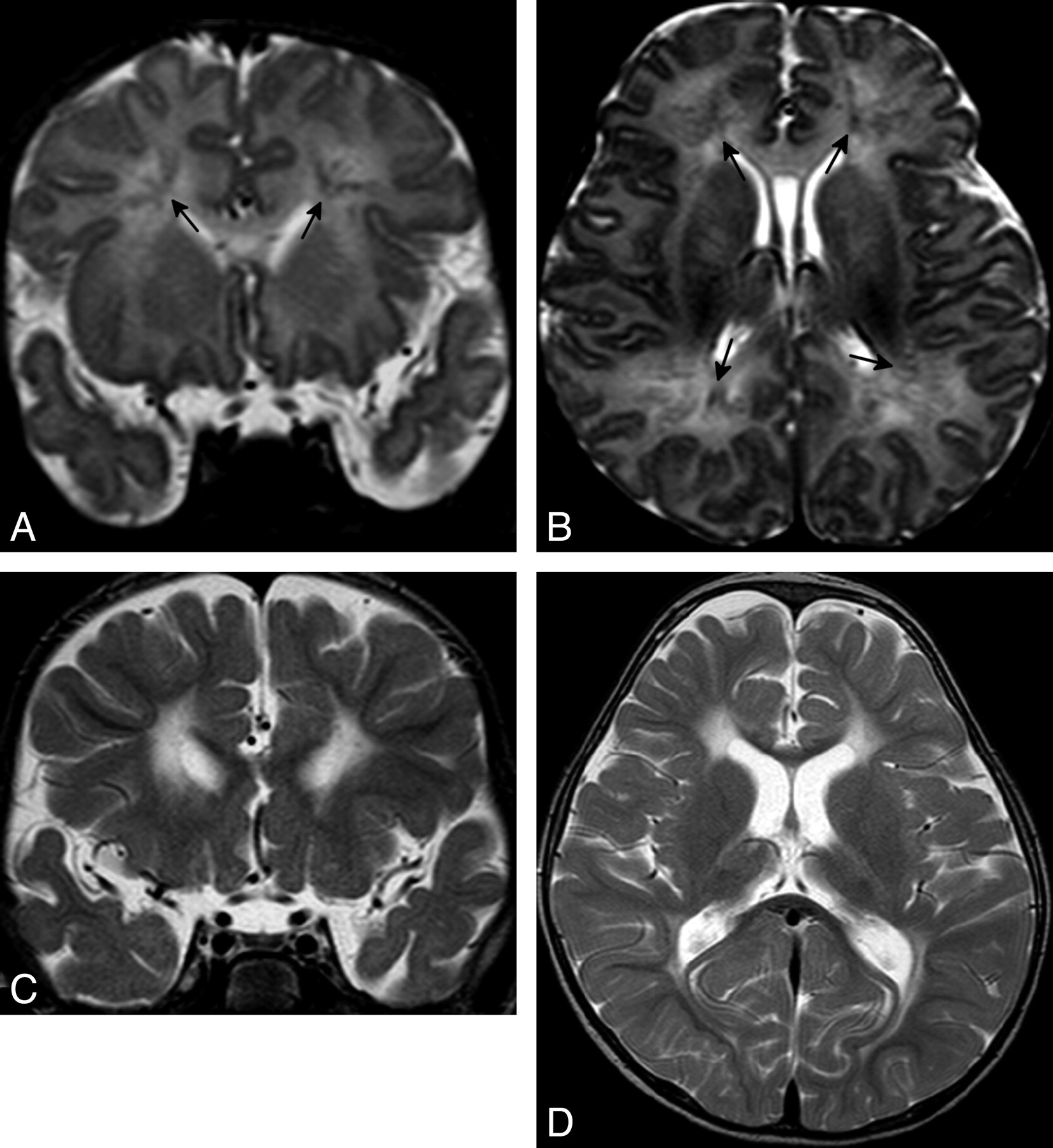
A–C: Axial T 2 -weighted magnetic resonance (MR) images revealing ...
(PDF) mTOR Pathway Mutations Cause Hemimegalencephaly and Focal ...
HHV-7–related encephalitis in 2-year-old girl with febrile convulsions ...
Neuroimaging In Cockayne Syndrome | American Journal of Neuroradiology
Macrocephaly, epilepsy and intracranial cysts: an image to remember ...
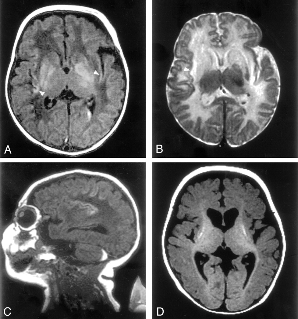
Brain MR Imaging in Neonatal Hyperammonemic Encephalopathy Resulting ...
Xanthoma Disseminatum: A Rare Intracranial Mass | American Journal of ...
Diffusion-Weighted Magnetic Resonance Imaging Identifies the ...
Phakomatoses: Tumor Suppression Gene Defects | Radiology Key
Infantile-onset Alexander disease in a child with long-term follow-up ...
MRI Brain: (a) bilateral T2 and FLAIR hyperintensities of lentiform ...
Refinement of cortical dysgeneses spectrum associated with TUBA1A ...
(PDF) Investigations of Huntington’s Disease and Huntington’s Disease ...
Brain MRI tumor classification. a Axial T2-weighted turbo spin-echo MRI ...
Brain MRI of patients 1 ( A and B ) and 3 ( C and D ). ( A ...
New Syndrome Characterized by Hypomyelination with Atrophy of the Basal ...
Brain MRI. ( A) Axial T2-weighted image of the brain of patient 1 ...
Focal splenial hyperintensity in epilepsy | Journal of Neurology ...
Nipah Viral Encephalitis or Japanese Encephalitis? MR Findings in a New ...
MRI of the brain showing right sided hemimegalencephaly and pachygyria ...
A 15-year-old boy with a 22month history of SSPE, clinical stage ...
Pontine ischemic rarefaction due to état criblé, white matter lesions ...
Traumatic Brain Injury: Diffusion-Weighted MR Imaging Findings ...
Restricted Diffusion in Vanishing White Matter | JAMA Neurology | JAMA ...
MR Imaging of Cerebral Cortical Involvement in Aceruloplasminemia ...
HSV-2 encephalitis. Sequential axial FLAIR images ( A – D ) show patchy ...
Deep Medullary Vein Involvement in Neonates with Brain Damage: An MR ...
MR Imaging with Diffusion-Weighted Imaging in Acute and Chronic ...
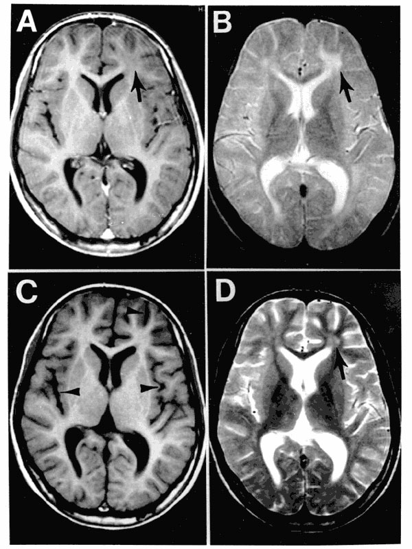
Figure 3 from MR imaging of the brain in Wilson disease of childhood ...
[Case 3]: Brain MRI of a 25-year-old male diagnosed with dengue fever ...
MR Imaging with Diffusion-Weighted Imaging in Acute and Chronic ...
T1-weighted-MRI
Normal-Brain-MRI-T2
Normal-Brain-MRI-with-Contrast
Abnormal-Brain-MRI-without-Contrast
Normal-Head-MRI-with-Contrast
Normal-Brain-MRI-Coronal
Normal-Brain-MRI-T1-Axial
MRI-T1-T2-Flair
Normal-Brain-MRI-Labeled
Female-Normal-MRI-Brain
Normal-Brain-Stem-MRI
MRI-Brain-Scan
Blood-MRI-T1-T2
Human-Brain-MRI
Normal-MRI-Brain-Pituitary
Normal-Brain-MRI-Side-View


![[Case 3]: Brain MRI of a 25-year-old male diagnosed with dengue fever ...](https://www.researchgate.net/publication/365860380/figure/fig4/AS:11431281103778618@1669815527426/Case-3-Brain-MRI-of-a-25-year-old-male-diagnosed-with-dengue-fever-showing-a.png)
