Please enter url.
Login
Logout
Please enter url.
Patient 3: Cavity, Edema, and Secondary Tumor Simulation, Fraction 1 ...
researchgate.net
source
Comments
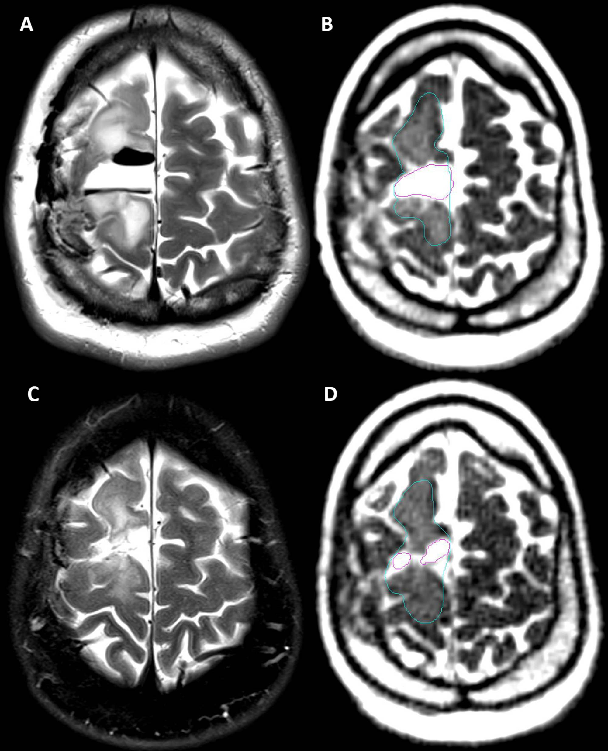
Table 1 from Daily Tracking of Glioblastoma Resection Cavity, Cerebral ...
Cureus | Daily Tracking of Glioblastoma Resection Cavity, Cerebral ...
Understanding White Matter Disease | Stroke
Chest X-ray and two different brain CT examinations of Patient 3 a-d ...
Hypomyelinating leukodystrophies: Translational research progress and ...
Diffusion MR Imaging of Hypoglycemic Encephalopathy | American Journal ...
Acute encephalopathy with biphasic seizures and late reduced diffusion ...
Diffusion Imaging in Brain Infections | Radiology Key
Stroke-Like Migraine Attacks After Radiation Therapy Syndrome | Ochsner ...
Figure 1 from Tissue-Specific Imaging Is a Robust Methodology to ...
Tissue-Specific Imaging Is a Robust Methodology to Differentiate In ...
Conventional MR scans show the appearance of MS lesions in the brain ...
Patient 1: a) FLAIR-weighted image showing hyperintense lesions on ...
MRI on Fluid-attenuated inversion recovery (FLAIR) and... | Download ...
Axial FLAIR reformats in patients without (A) and with imaging features ...
Lesions of seizure induced brain edema in a 64-year-old male. MRI and ...
Frontiers | A Method to Experimentally Estimate the Conductivity of ...
Diagnostic Value of 18F-FDG PET/CT Versus MRI in the Setting of ...
Imaging and CSF analyses effectively distinguish CJD from its mimics ...
Posttransplant lymphoproliferative disease (PTLD): a patient with a ...
Delayed Neurotoxicity in Primary Central Nervous System Lymphoma ...
Pediatric Acute Toxic Leukoencephalopathy: Prediction of the Clinical ...
Figure 1 from Usefulness of diffusion tensor imaging of white matter in ...
Diagnostic approach in a patient with Creutzfeldt-Jakob disease ...
SCN8A Epileptic Encephalopathy: Detection of Fetal Seizures Guides ...
The shear-strain effects of impact head injury are shown in the case ...
Posterior reversible encephalopathy syndrome (PRES) in a patient with ...
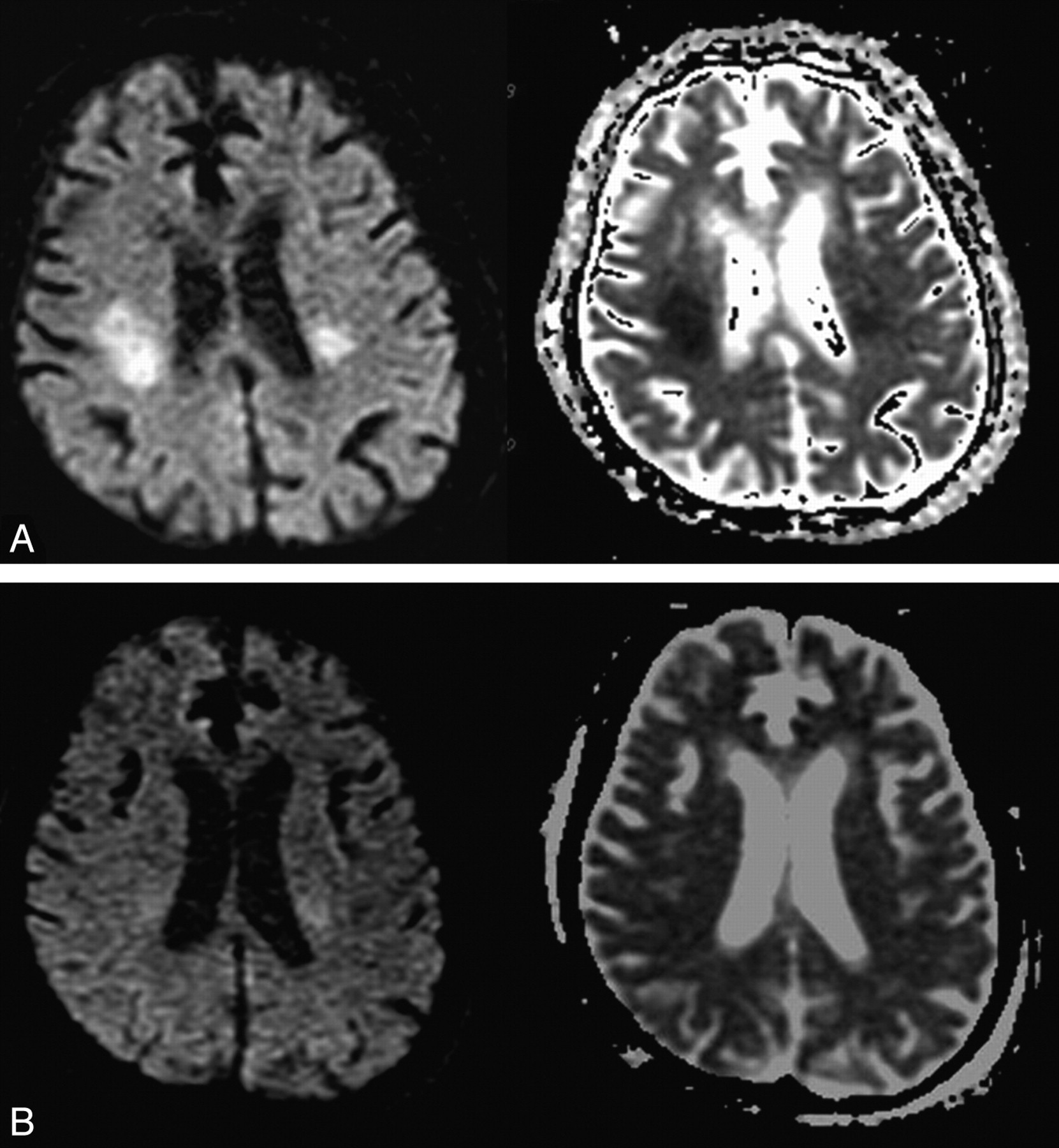
Utility of diffusion-weighted imaging (DWI) and apparent diffusion ...
On day 1, Symmetrical hyper-intensity lesions on DWI-MRI (A) over ...
SVR technique performed on a fetus at 33 weeks of GA. Three orthogonal ...
First CT Findings (done within 2 h from complaint starting): showing ...
Figure 1 from Imagerie de diffusion et maladie de Creutzfeldt-Jakob ...
Figure.Brain magnetic resonance image (day 20 after admission in ...
FDG-PET images of a pineocytoma, b PPTID, c pineoblastoma, and d ...
The b0 of the DWI (a) of a 77 years old male showed WMH (Fazekas scale ...