Please enter url.
Login
Logout
Please enter url.
Right ventricular enlargement in transthoracic echocardiography ...
researchgate.net
source
Comments
Right ventricular enlargement in transthoracic echocardiography ...
Decreased TAPSE in transthoracic echocar diography. | Download ...
GE Venue auto ivc - YouTube
Parasternal short axis view shows high velocity continuous flow of ...
SPI-Display Modes Flashcards | Quizlet
Masquerading Bundle Branch Block in a Patient with Heart Failure in Ni ...
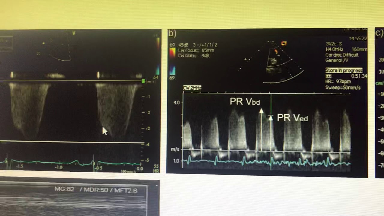
PAEDP and mean PAP by echocardiogram - YouTube
Enlarged image of right ventricle. Small amount of pericardial effusion ...
Parasternal long-axis view showing normal LV function. | Download ...
Congenital Heart Defect Stock Photos, Pictures & Royalty-Free Images ...
Intravenous Immunoglobulins in the Management of Parvovirus B19 Induced ...
Intrahepatic portosystemic venous shunt: colour Doppler and MDCT ...
Transthoracic echocardiography in M mode and parasternal view, showing ...
Prague ICU
(PDF) Internal jugular vein distensibility in assessment of fluid ...
Low TAPSE value (15 mm) in first echocardiography. TAPSE, tricuspid ...
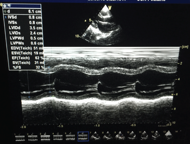
Echocardiographic image showed ejection fraction of left ventricle. The ...
M mode of echocardiograph showing increased gradient across aortic ...
M mode in aortic stenosis - YouTube
SciELO - Brasil - Preoperative assessment of inferior vena cava ...
Cardiac ultrasound results. (a) Results obtained upon admission to the ...
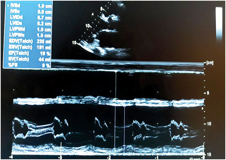
Echocardiography showed features of dilated cardiomyopathy. | Download ...
Parasternal short axis view shows high velocity continuous flow of ...
Heart: dilated cardiomyopathy (DCM) in Dogs (Canis) | Vetlexicon
This figure shows M-mode echocardiogram of left atrium and aortic root ...
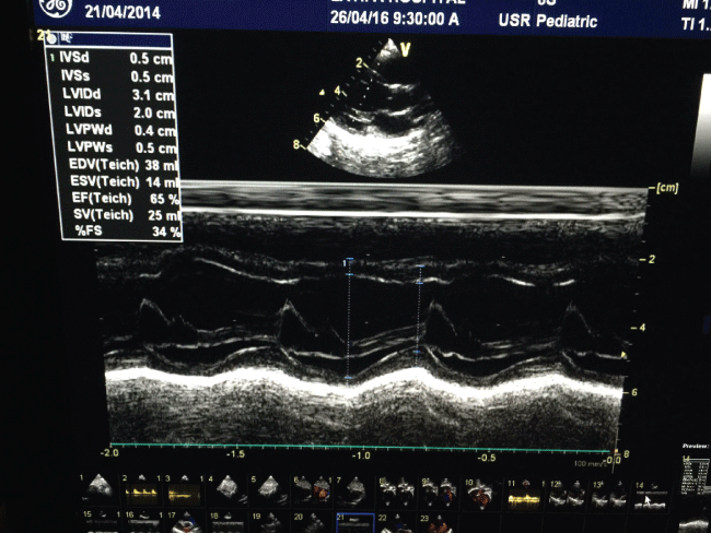
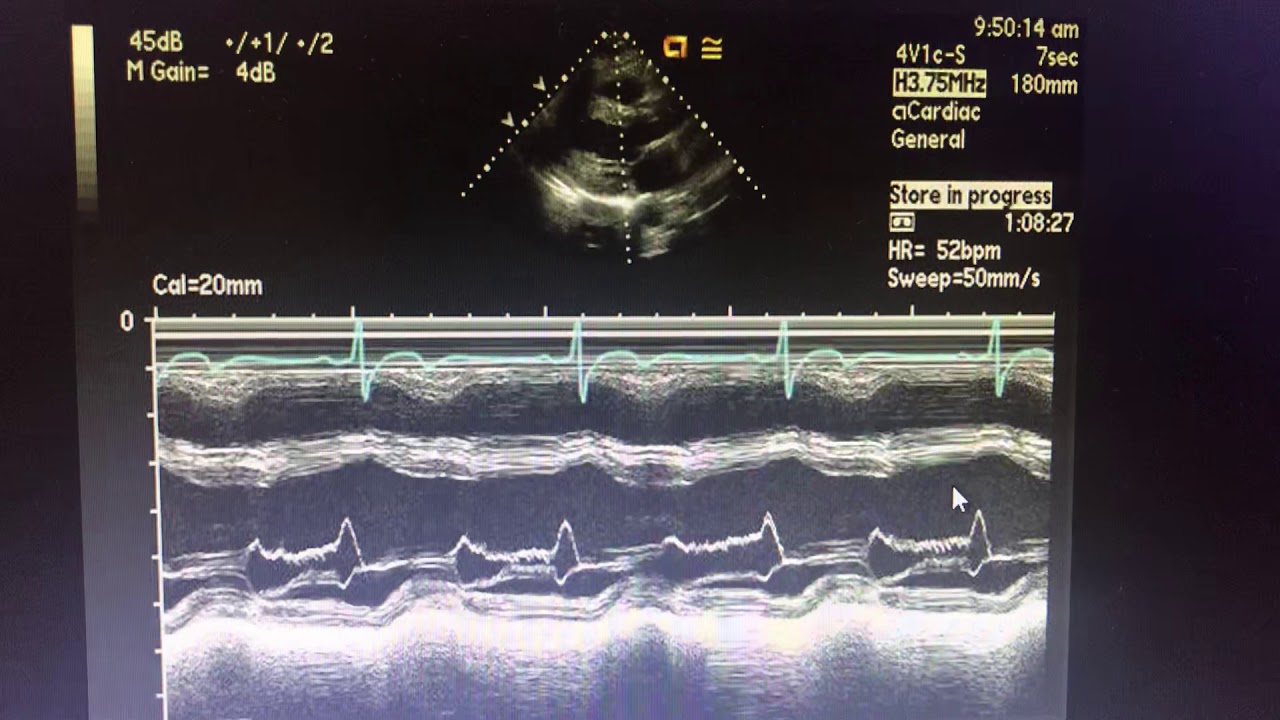
M-mode guided by 2-D echocardiography for LV EF% calculation ...
Value of duplex doppler ultrasonography in non-invasive assessment of ...
Intravenous Immunoglobulins in the Management of Parvovirus B19 Induced ...
Intrahepatic portosystemic venous shunt: colour Doppler and MDCT ...
Minimal pelvicalyceal dilatation is seen on gray scale US in the left ...
M mode in aortic regurgitation - YouTube
M-mode view of the heart. | Download Scientific Diagram
Echocardiography in Noncardiac Surgery | Thoracic Key
Doppler Echocardiography showing restrictive pattern of diastolic ...
Doppler ultrasound in obstetrics - Obstetrics, Gynaecology and ...