Please enter url.
Login
Logout
Please enter url.
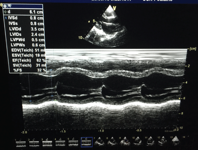
Transthoracic echocardiography in M mode and parasternal view, showing ...
researchgate.net
source
Comments
The M Mod Echocardiogram show systolic disfunction during the last ...
Professional Sonoscape E1 Veterinary Ecografo Sonoscape Brand - Buy E1 ...
Enlarged image of right ventricle. Small amount of pericardial effusion ...
Intravenous Immunoglobulins in the Management of Parvovirus B19 Induced ...
(PDF) Left ventricular subvalvar aneurysm with severe mitral ...
Used Very Good HITACHI EUP-S72 ID#1936189 for Sale item# 1936189 | Bimedis
Brain MRI T2-weighted images showing hyperintense lesions in ...
Passive hepatic congestion: Findings of two-dimensional, Doppler, and ...
Tissue Doppler echocardiography velocity profile at septal mitral ...
Transthoracic echocardiography in M mode and parasternal view, showing ...
echocardiogram:multiple rhabdomyoma tumor. cw doppler - YouTube
The echocardiographic examination revealing the pericardial effusion in ...
Monitoring of Aortic Valve Opening and Systolic Aortic Insufficiency in ...
Cyclic variation in tricuspid inflow was significantly improved from ...
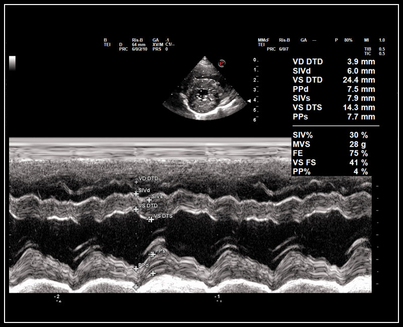
PLAX view showing M-mode across left ventricle. | Download Scientific ...
Colour Doppler of ovarian mass showing a high peak systolic velocity of ...
Echocardiographic findings showing severe Tricuspid regurgitation ...
Chapter 15 - Echocardiography Board Review: 500 Multiple Choice ...
VET Ultrasound Clinical Images
DOPPLER ULTRASOUND_normal versus reduced inferior vena cava modulation ...
E: M-mode from right parasternal long axis view depicting systolic ...
TRANSDUCERS, IMAGE FORMATION, AND ARTIFACTS | Anesthesia Key
Figure 5 from A novel technique for invasive aortic valve pressure ...
Echocardiographic image of pulsed wave Doppler spectral display of flow ...
Transthoracic Doppler echocardiogram showing ejection fraction of 37% ...
VET Systems
Monitoring of Aortic Valve Opening and Systolic Aortic Insufficiency in ...
Transesophageal echocardiography (TEE) fi ndings: four-chamber view ...
Left ventricular free wall systole (LVFWs) and left ventricular free ...
SonoZone: Ultrasound Modes: A, B, & M Sonography, Ultrasound, Echo, Work
Doppler ultrasound in obstetrics - Obstetrics, Gynaecology and ...
M Mode view of the IVC in a ventilated patient demonstrating 23% ...
22 Weeks - Braxton Hicks - NEW ULTRASOUND
Echo IV | Anesthesia Key
Echocardiograms in 2D and M-mode during induction of AICM ...