Please enter url.
Login
Logout
Please enter url.
Terri Schiavo Grave
ar.inspiredpencil.com
source
Comments
*Evil under the Sun*: August 2008
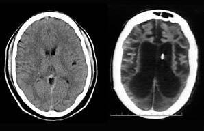
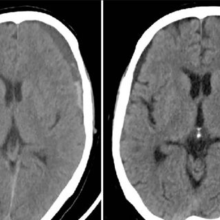
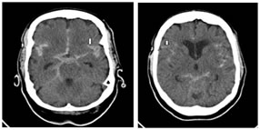
Noncontrast CT scan and MRI of the brain with DWI
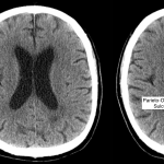
Radiological Anatomy: Parieto-Occipital Sulcus - Stepwards
Frontiers | A Critical Review of Alberta Stroke Program Early CT Score ...
Radiological Anatomy: Parieto-Occipital Sulcus - Stepwards
(a and b): CT scan of the brain axial views showing multiple lytic ...
Don't Try This Alone: Silent Epidemic of Attachment Disorder
Always in Tune: The Unforgettable Memory for Music in Alzheimer’s ...
(PDF) Contrast-induced encephalopathy after coronary angioplasty and ...
Malignant Glioma: Symptoms, Diagnosis and Treatment - Symptoma®
Dr Balaji Anvekar FRCR: 01/11/2011 - 01/12/2011
Radiological Anatomy: Parieto-Occipital Sulcus - Stepwards
Role of Endoscopic Third Ventriculostomy and Ventriculoperitoneal Shunt ...
A 50-year-old female patient who has sulcal hyperintensity on ...
Frontiers | Anatomical and Physiological Differences between Children ...
(a) Initial CT scan showing left CSDH and (b) CT scan after irrigation ...
(PDF) Usefulness of embolization of the middle meningeal artery for ...
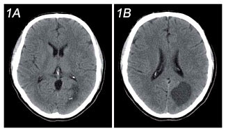
(a and b) Computed tomography brain showing a chronic subdural hematoma ...
Pseudotumor Cerebri vs Hydrocephalus; Is There a Difference? - Med ...
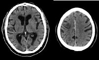
CT scans, showing C.F.'s lesion. Ischemic areas appear darker as ...
Descending transtentorial herniation. A right temporal large traumatic ...
Radiology MRI: Acute MCA Infarct on CT
Degenerative and Demyelinating Diseases | Radiology Key
CT scans of the brain at SLE diagnosis (A) and current presentation (B ...
Demonstrates the change in volume of depressed brain over time. Volume ...
(PDF) Acute parietal lobe infarction presenting as Gerstmann’s syndrome ...
SIHE: After falling from standing position, CT images within 3 hours ...
Resonancia magnética cerebral al decimocuarto día de ingreso: área ...
MBBS Medicine (Humanity First): CT SCAN OF HEAD
(PDF) Chronic Subdural Hematoma Infected by Propionibacterium Acnes: A ...
Volume 17 Issue 3
CT scans of the brain at SLE diagnosis (A) and current presentation (B ...
Left Sylvian artery stroke. (A) Non-contrast CT weighted sum image ...
Neuro Image December 2005 Page 3
CT scan of DNR. Axial views of the CT scan show the extensive lesion in ...
Brain-Damage-CT-Scan
Abnormal-Brain-CT
Abnormal-Head-CT-Scan
Human-Brain-CT-Scan
Stroke-CT-Brain-Scan
Traumatic-Brain-Injury-CT-Scan
Cat-Scan-Brain
Healthy-Brain-CT-Scan
CT-Scan-Brain-Lobes
Brain-CT-Scan-with-Contrast
Head-and-Neck-CT-Scan
CT-Brain-3D
MRI-Brain-Scan
Brain-Cancer-CT-Scan
Brain-CT-Scanner
Empty-Brain-Scan