Please enter url.
Login
Logout
Please enter url.
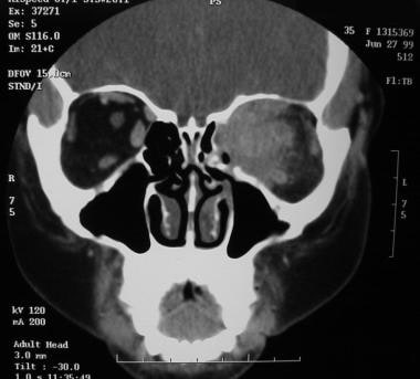
Figure 2 from Treating nevoid basal cell carcinoma syndrome. | Semantic ...
semanticscholar.org
source
Comments
Neoplasia and Tumorlike Lesions | Ento Key
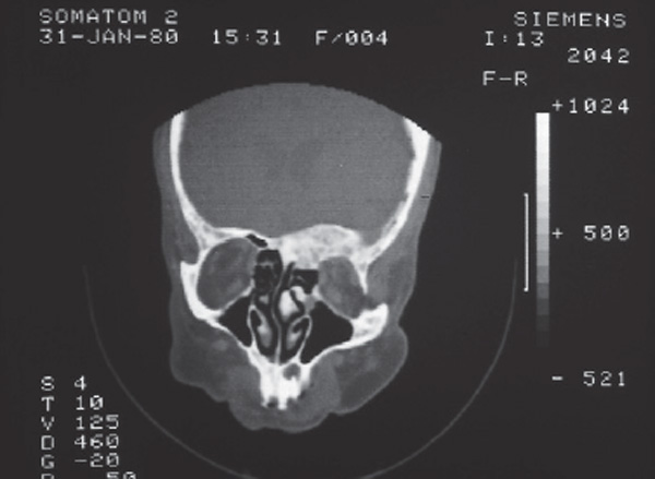
Figure 1 from Suppurative osteomyelitis Non suppurative osteomyelitis ...
Tomografía computarizada simple para ventana ósea que muestra ...
Surgical treatment of choanal atresia with transnasal endoscopic ...
Tumors of the Lacrimal Drainage System | Ento Key
An Introductory Guide to Sinonasal Tumors. #nasalobstruction # ...
otolaryngology-Left-side
Fibro-osseus lesions Flashcards | Quizlet
B: Coronal and axial views showing the lesion with obliteration of ...
Transverse section of computerized tomography orbits showing bilateral ...
CT in coronal view: absence of middle and inferior left turbinates ...
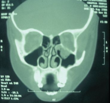
CT showing the tumour in right maxillary sinus. | Download Scientific ...
Exposure with suprastructure maxillary swing | Download Scientific Diagram
TC que evidenció colección sin aparente conexión con estructuras ...
CECT of PNS (axial view) | Download Scientific Diagram
Preoperative anterior view of the patient showing left eye ptosis ...
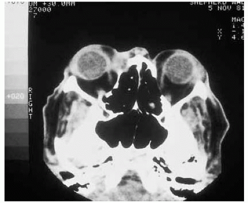
Axial CT following intravenous contrast showing bilateral orbital ...
Intraosseous ancient Schwannoma: A rare case in the mandible and a ...
Figure 1 from Prognostic value of sinus CT scans in hematopoietic stem ...
Figure 1 from Retinal Astrocytoma Associated with Hypertrophy of the ...
CT scan presentation. Solid tumor full filled the left orbit Fig. 3 ...
The Anatomic Relevance of the Haller Cell in Sinusitis | Semantic Scholar
10 Maxillofacial surgery | Pocket Dentistry
Rhinocerebral mucormycosis: A case of a rare, but deadly disease ...
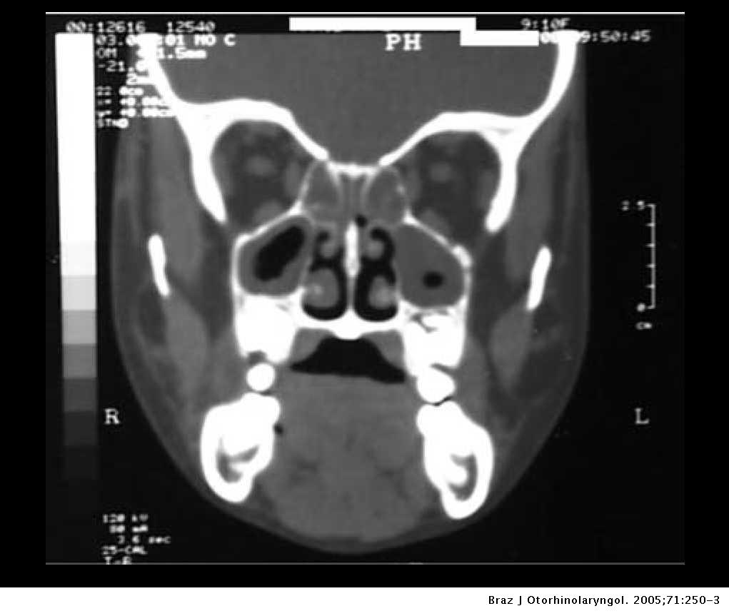
Rhinosinusitis in a patient with Behçet's syndrome | Brazilian Journal ...
Figure 1 from Midline Maxillary Odontogenic Keratocyst | Semantic Scholar
(PDF) Pneumatization of the sphenoid sinus
Figure 1 from Asymptomatic antrolith in maxillary sinus. Report of a ...
Computed tomography scan confirming the clinical suspicion of extrusion ...
Coronal CT showing a heterodense soft tissue mass in the right ...
Orbit Anatomy: Osteology, Lacrimal System, Connective Tissue Planes
b. Bone window setting of the zygoma-infraorbital margins showing ...
Hydatid cyst of the parotid gland - دکتر علیرضا محبی
Clinical photograph showing features of right exophthalmos and left ...
Corte axial de TC cérvicofacial que muestra el engrosamiento de partes ...