Please enter url.
Login
Logout
Please enter url.
Figure 1 from Midline Maxillary Odontogenic Keratocyst | Semantic Scholar
semanticscholar.org
source
Comments
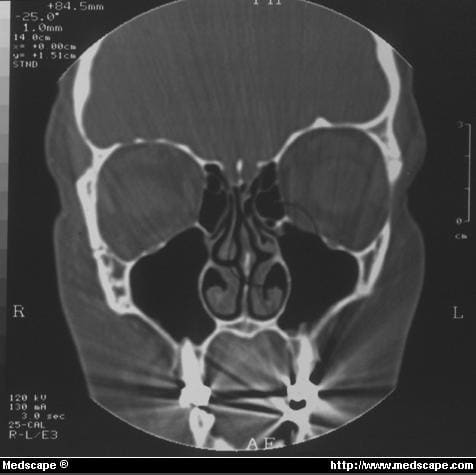
A Coronal CT section (patient 6) at level of midportion of left globe ...
Figure 1 from Maxillary Sinus Involution after Endoscopic Sinus Surgery ...
CECT scan of nose and PNS—showing contrast enhancing mass in the right ...
Contrast enhanced CT (coronal section) of paranasal sinuses showing ...
The Anatomic Relevance of the Haller Cell in Sinusitis | Semantic Scholar
Coronal CT scan demonstrates mucosal thickening, air-fluid levels and ...
Preoperative sinus computed tomography. The figure shows a piece of ...
Classification of fungal sinusitis | Download Scientific Diagram
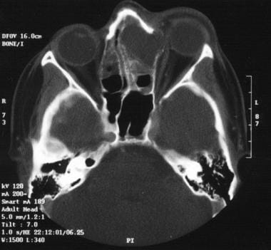
Postoperative axial bone algorithm CT at level of sphenoid sinus (arrow ...
Photograph shows the viable right middle turbinate after it was removed ...
(PDF) Traumatic Intracranial Frontal Extradural Hematoma Associated ...
Patient 4 one day after water balloon injury showing marked ecchymoses ...
Computed tomography scan showing radiolucent-radiopaque mass in the ...
Functional Endoscopic Sinus Surgery for Sinusitis - Page 3
Figure 1 from Complication of maxillary sinus Foley balloon placement ...
Coronal (A) and axial (B) sections of Computed tomography of a patient ...
e 2. Tomografia de seios paranasais, corte axial, janela para partes ...
Figure 1 from Middle Meatal Antrostomy Stenting following Pediatric ...
Internet Scientific Publications
A PS (posteriosuperior) deviation is seen. Deviation is located in the ...
Preoperative CT scan of axial section of mid-orbit shows large mucocele ...
CT scan of nose and paranasal sinuses showing diffuse mucosal ...
CT scan of paranasal sinuses, coronal view showing an extensive mass ...
Corte orbitario de la tomografía de cráneo. No se encontraron ...
Infections of the Lacrimal Drainage System | Ento Key
Figure 1 from Role of Haller's Cell in Headache and Sinus Disease: A ...
(A) Peripheral blood smear (Wright-Giemsa stain; original... | Download ...
Tomografia computadorizada. | Download Scientific Diagram
Radiographic imaging studies in pediatric chronic sinusitis - Journal ...
CT scan of the orbits, coronal view, demonstrating gross enlargement of ...
(PDF) Bilateral Massive Conchae Bullosa Mimicking Intranasal Tumors
Surgical Treatment of Acute Maxillary Sinusitis: Overview, Surgery for ...
The Anatomic Relevance of the Haller Cell in Sinusitis | Semantic Scholar
Coronal view of the CT scan of the paranasal sinus showing the osteoma ...
Orbitoethmoid Osteoma: Case Report of an Uncommon Presentation of an ...