Please enter url.
Login
Logout
Please enter url.
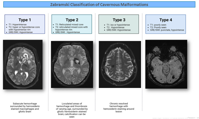
Figure 1. [Zabramski classification of cavernous malformations ...
ncbi.nlm.nih.gov
source
Comments
Additional distinguishing MRI features between cerebral small vessel ...
DYKE-DAVIDOFF-MASSON SYNDROME-A Rare Cause of Cerebral Hemiatrophy in a ...
Review of Radiologic Considerations in an Immunocompetent Patient With ...
Chronic dysfunction of blood-brain barrier in patients with post ...
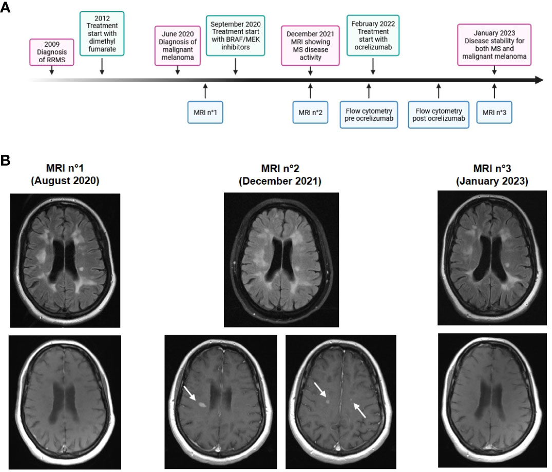
Frontiers | Case Report: Balancing immune responses – multiple ...
Brain atrophy progression before gene therapy. Representative MRI ...
EPOS™
Schematic Overview of Image Processing. Note: This schematic overview ...
Mitochondrial Encephalomyopathy With Lactic Acidosis and Strokelike ...
A Case of CNS Nocardia farcinica Presenting as Aphasia in an ...
A 64-year-old male with recurrent glioblastoma shows heterogeneous ...
Cardinal MRI difference between cerebral small vessel disease and ...
Ilustrative case of right frontal oligodendroglioma, primary case. A ...
Treatment of natalizumab‐associated PML with filgrastim - Stefoski ...
Comparison of Boston Criteria v2.0/v1.5 for Cerebral Amyloid Angiopathy ...
MRI phenotype 5-year conversion rates | Download Scientific Diagram
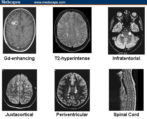
Multiple Sclerosis: Diagnosis and Management Strategies
(PDF) Comparison of MRI and CT for Detection of Acute Intracerebral ...
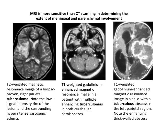
Cns infections radiology.
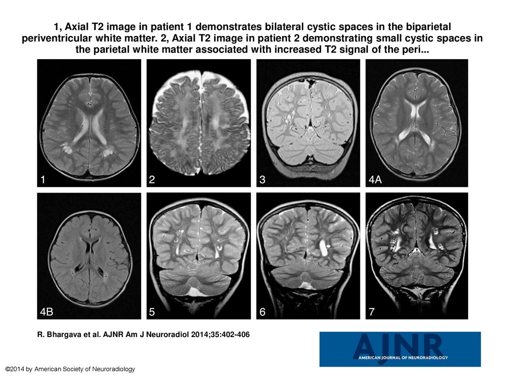
1, Axial T2 image in patient 1 demonstrates bilateral cystic spaces in ...
Creutzfeldt-Jakob Disease - Diffusion weighted and diffusion tensor ...
MRI findings in patients with MOG-associated encephalomyelitis. a1-a3 ...
Figure 2 from MRI Characteristics of Autoimmune Encephalitis With ...
Can You See Alzheimers On A Ct Scan - Ellie Matthew's Blog
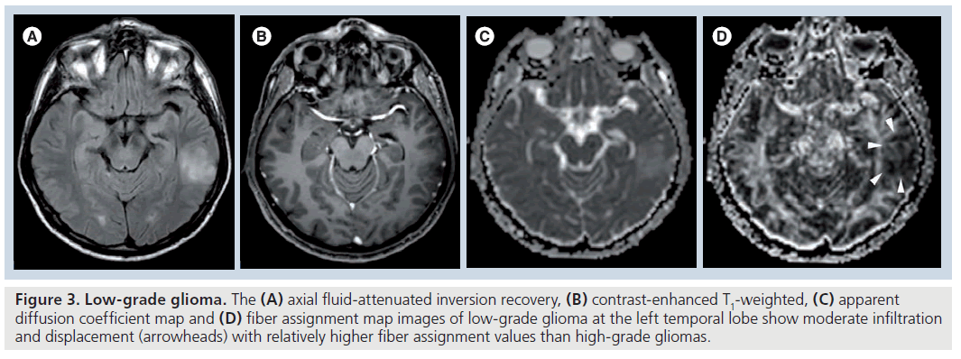
Diffusion-tensor imaging in brain tumors
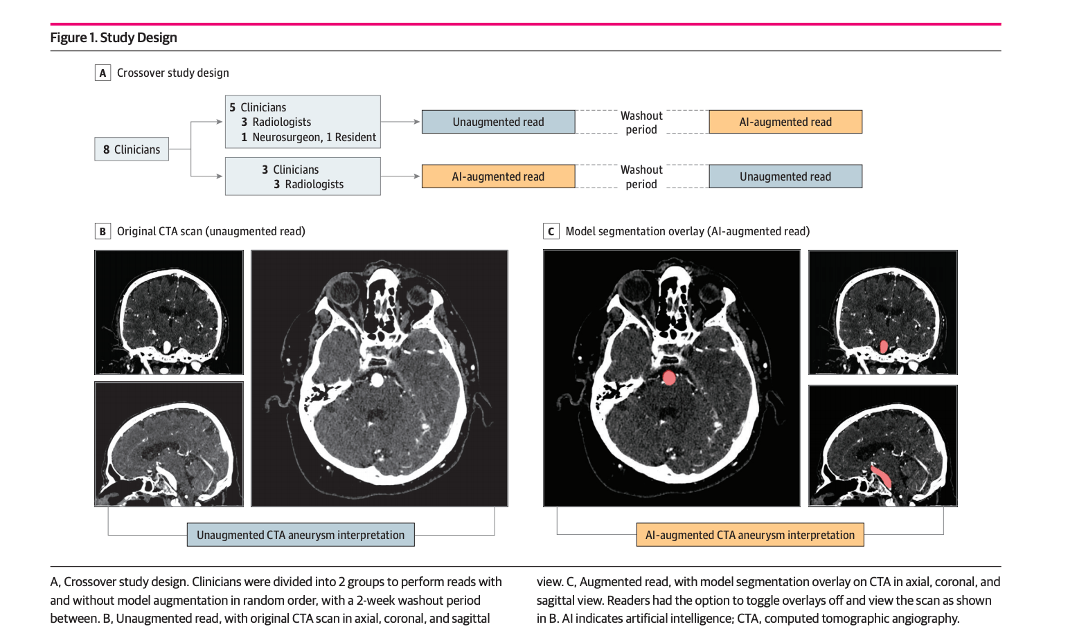
Deep Learning–Assisted Diagnosis of Cerebral Aneurysms | Leaders in ...
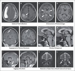
Spontaneous intracranial hypotension
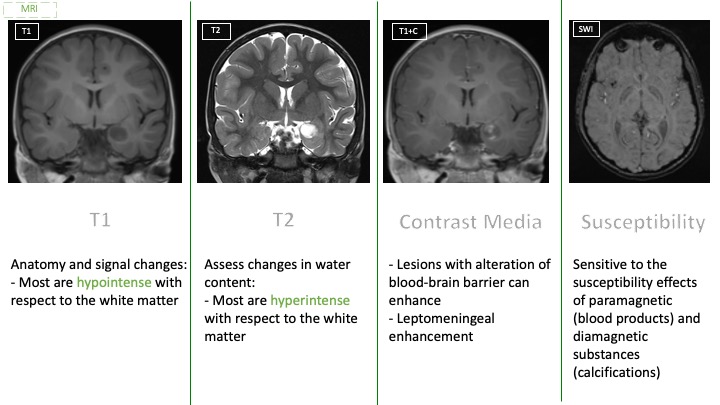
Introduction to mri
Examples of acute cerebral microembolism after atrial fibrillation ...
Magnetic resonance imaging scans of the patient’s brain. Images show ...
Lesion location on T2-weighted MR scans. Axial T2-weighted images ...
Example of a positive hyperdense lesion patient. The patient with ...
50 PML in a 67-year-old male with a history of diffuse large B-cell ...
Imaging in pediatric brain tumors