Please enter url.
Login
Logout
Please enter url.
Brain atrophy progression before gene therapy. Representative MRI ...
researchgate.net
source
Comments
Long-Term Follow-Up After Gene Therapy for Canavan Disease | Science ...
Pallidoreticular Damage in Acute Carbon Monoxide Poisoning: Diffusion ...
Follow-up MR images after five months in comparison with previous MR ...
Oral cysteamine bitartrate and N-acetylcysteine for patients with ...
Figure 1 from CADASIL presenting with a behavioural variant ...
Fusion results of MRT1/ MRT2 images. a MRT1 image, b MRT2 image, c ...
The protean manifestations of central nervous system IgG4-related ...
Brain atrophy progression before gene therapy. Representative MRI ...
Magnetic resonance imaging (MRI) and computerized tomography ...
Representative longitudinal T 1 and T 2 weighted scans in a neonate, 1 ...
Pseudo-Subarachnoid Hemorrhage due to Leptomeningeal Carcinomatosis
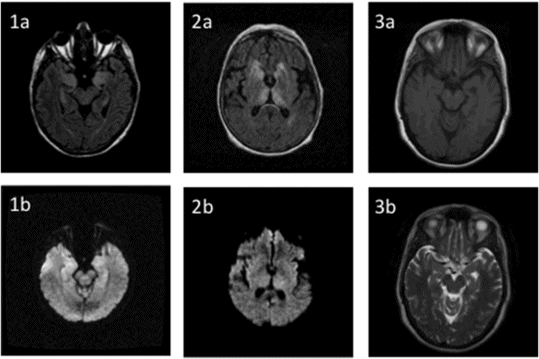
1 Brain MR (FLAIR and T2*-weighted images) of patient A showing a ...
Stress‐Induced Upregulation of SLC19A3 is Impaired in Biotin‐Thiamine ...
Samples of T2-weighted control and abnormal brain MR images [5 ...
First row CT image as fixed (left) slice 97, MR images as moving ...
Magnetic resonance fluid-attenuated inversion recovery (FLAIR) images ...
[PDF] TGF-beta1 is associated with the progression of intracranial deep ...
Test images of the Harvard Brain Atlas database | Download Scientific ...
Examples of different types of pathological brain. (a) normal; (b ...
(PDF) Traumatic brain injury and subsequent glioblastoma development ...
Sample testing, training, and different grade‐level MRI | Download ...
Brain MRI studies. T2-weighted (a–d) or susceptibility-weighted imaging ...
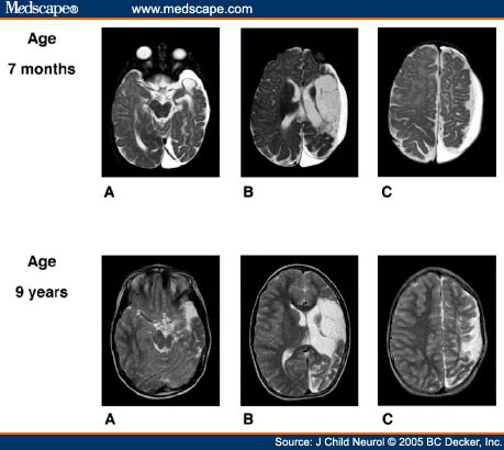
A 9-Year-Old Boy With a History of Large Perinatal Stroke - Page 2
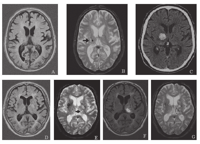
MRI brain axial FLAIR images (A-D) show hyperintense signal in right ...
Retinal vasculopathy with cerebral leukoencephalopathy and systemic ...
Applied Sciences | Free Full-Text | Connectivity Alterations in ...
Scans reveal how cocaine ‘eats away’ at your brain in a matter of ...
CMRI axial: Examples of different pathomorphologies and how GDAH was ...
| The brain magnetic resonance imaging (MRI) of the three patients with ...
MRI of the brain: (A) abnormal gyral hyperintensity in the left ...
Diffusion-weighted signal patterns of intracranial haemorrhage ...
Radiological progression of MAPT R406W-related neurodegeneration ...
Autoimmune antibody-associated encephalopathy and dementia syndromes ...
Effect of NOTCH3 EGFr Group, Sex, and Cardiovascular Risk Factors on ...
Persistent high-signal intensity lesions on diffusion-weighted imaging ...


![[PDF] TGF-beta1 is associated with the progression of intracranial deep ...](https://www.researchgate.net/profile/Toshiki-Mizuno/publication/257300020/figure/fig1/AS:443851438596098@1482833837342/Upper-Representative-deep-white-matter-lesion-DWL-cases-according-to-the-magnetic_Q320.jpg)