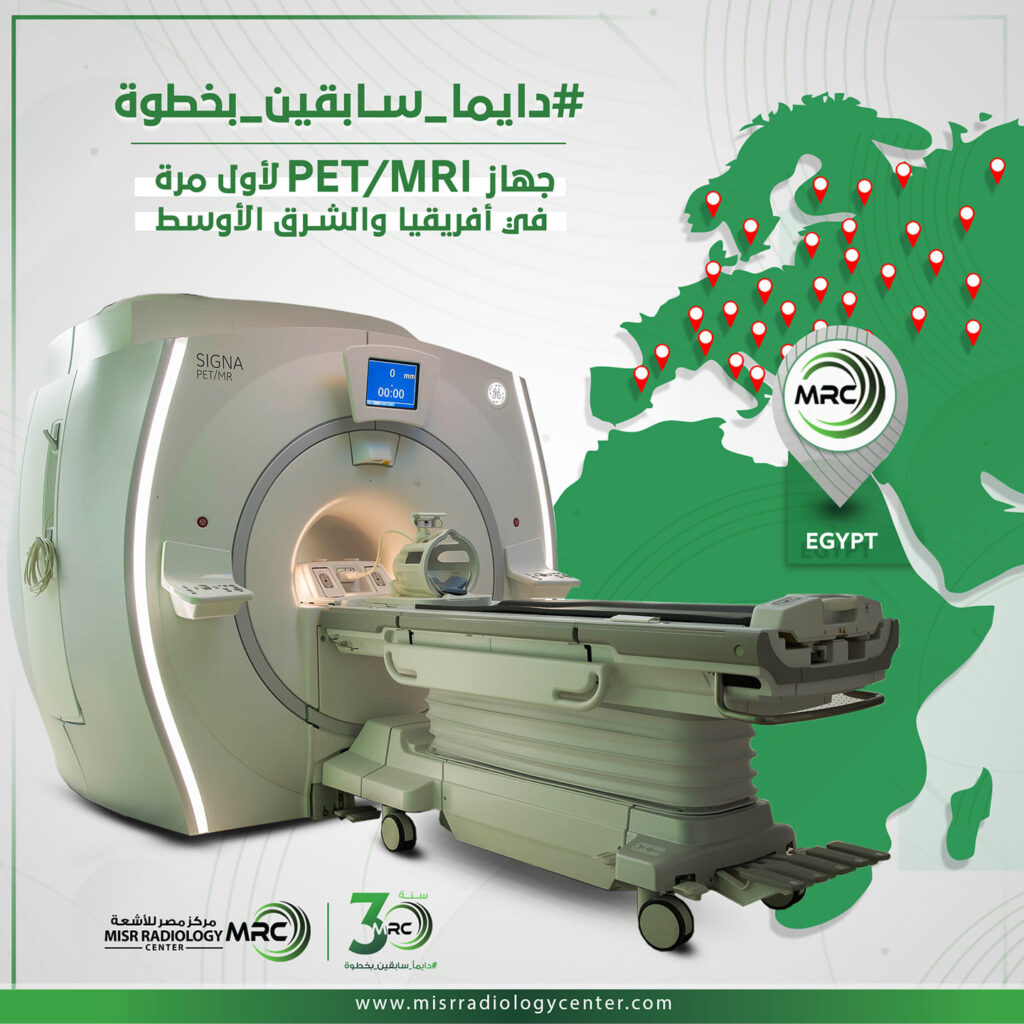
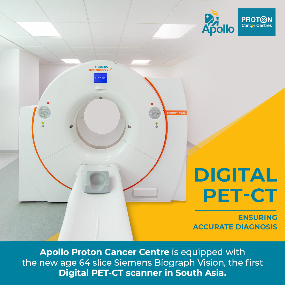
Apollo T 1st Pet/mri


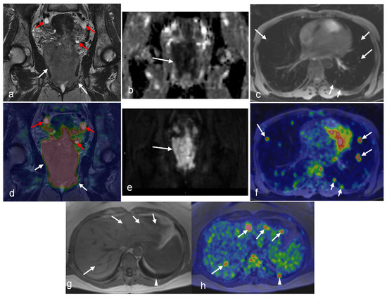


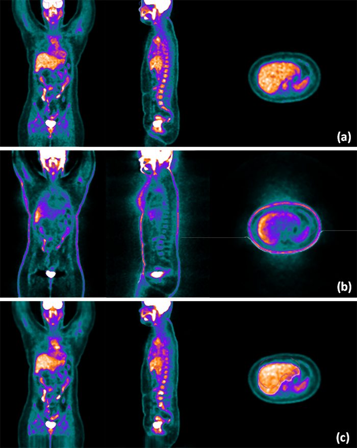
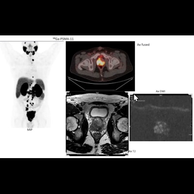
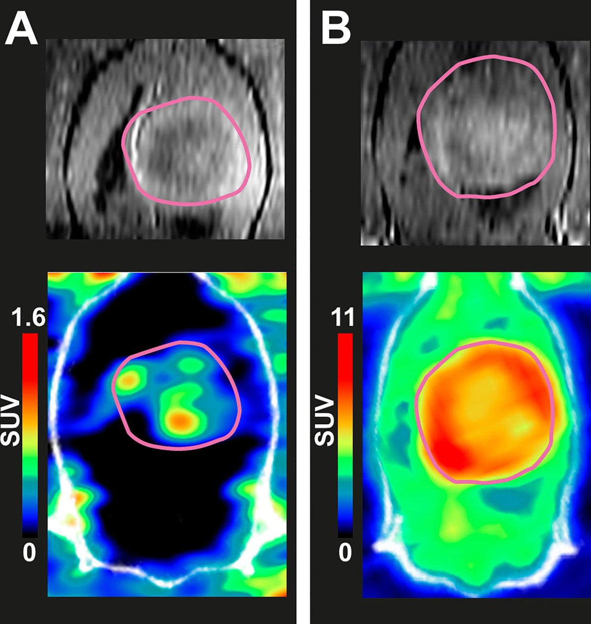
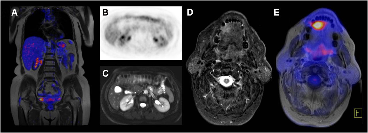
![A/C: Axial fused [ 89 Zr]Zr-Df-IAB22M2C PET/MRI and MRI of a metastasis ...](https://www.researchgate.net/publication/370540851/figure/fig1/AS:11431281155983243@1683303975990/A-C-Axial-fused-89-ZrZr-Df-IAB22M2C-PET-MRI-and-MRI-of-a-metastasis-with-intense_Q640.jpg)


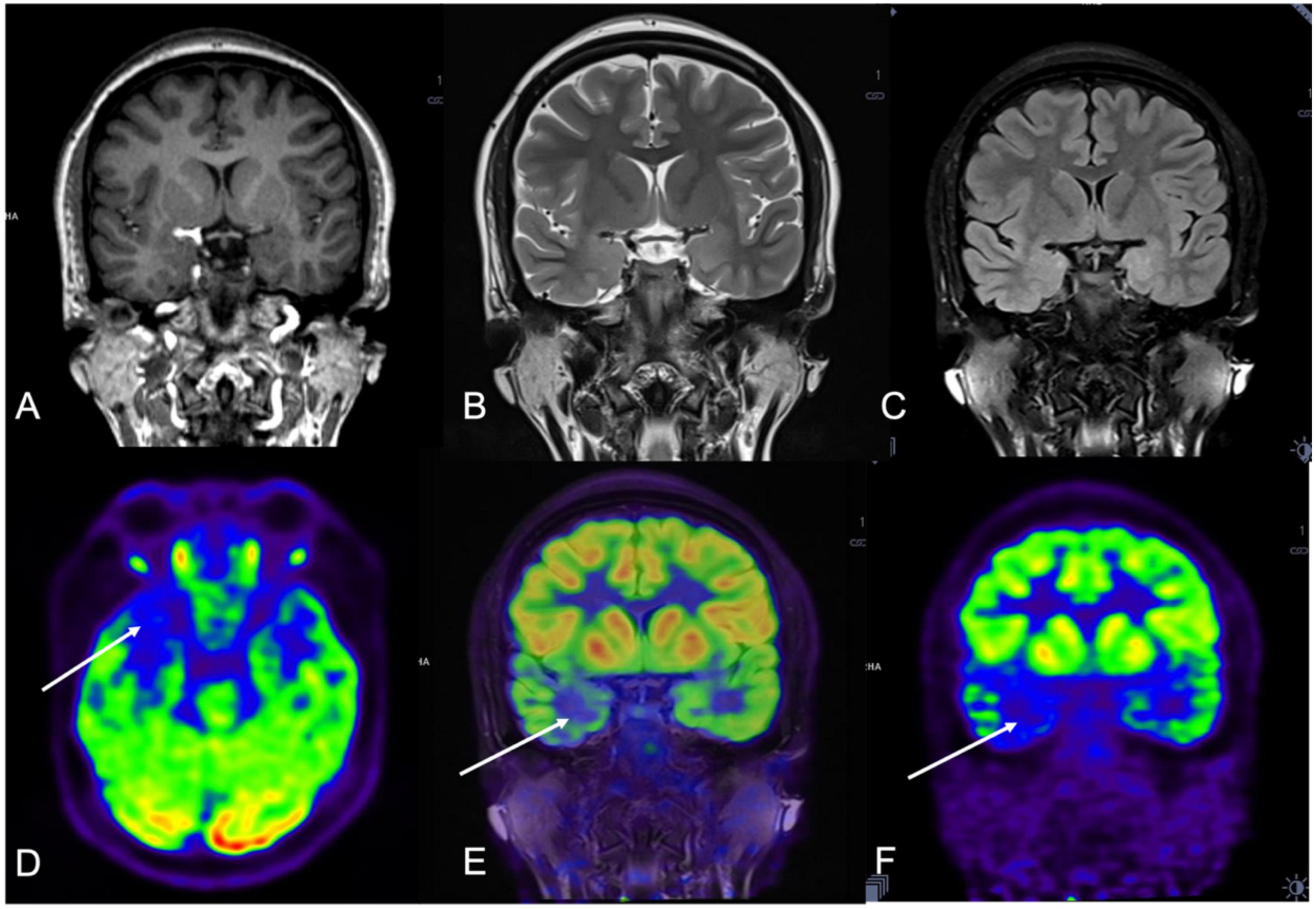


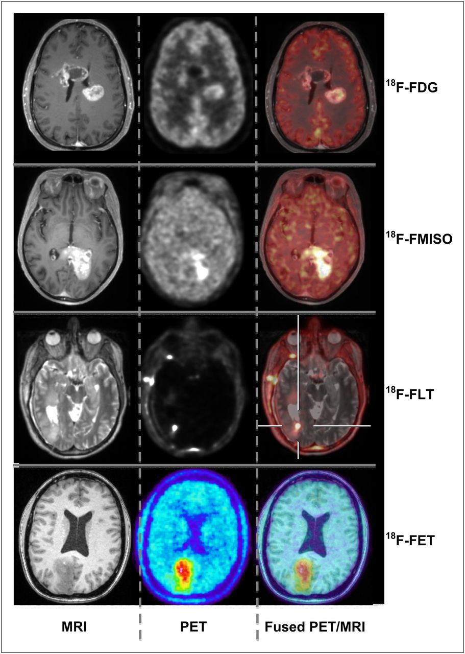
![MRI negative PET TLE. T1MR1 (left), [ 18 F] FDG-PET (right) and ...](https://www.researchgate.net/publication/8428197/figure/fig1/AS:281350125965312@1444090505250/MRI-negative-PET-TLE-T1MR1-left-18-F-FDG-PET-right-and-co-registered-MR1-and.png)


![MRI and [¹¹C]TMSX PET images of a healthy subject. Axial T1 ...](https://www.researchgate.net/publication/321192714/figure/fig2/AS:1086520727474178@1636058136217/MRI-and-CTMSX-PET-images-of-a-healthy-subject-Axial-T1-gadolinium-enhanced-weighted.jpg)


Experience the stunning modern approach to Apollo T 1st Pet/mri with comprehensive galleries of contemporary images. featuring the latest innovations in photography, images, and pictures. ideal for contemporary publications and media. Each Apollo T 1st Pet/mri image is carefully selected for superior visual impact and professional quality. Suitable for various applications including web design, social media, personal projects, and digital content creation All Apollo T 1st Pet/mri images are available in high resolution with professional-grade quality, optimized for both digital and print applications, and include comprehensive metadata for easy organization and usage. Our Apollo T 1st Pet/mri gallery offers diverse visual resources to bring your ideas to life. Advanced search capabilities make finding the perfect Apollo T 1st Pet/mri image effortless and efficient. Comprehensive tagging systems facilitate quick discovery of relevant Apollo T 1st Pet/mri content. Professional licensing options accommodate both commercial and educational usage requirements. Diverse style options within the Apollo T 1st Pet/mri collection suit various aesthetic preferences. Multiple resolution options ensure optimal performance across different platforms and applications. Whether for commercial projects or personal use, our Apollo T 1st Pet/mri collection delivers consistent excellence. Cost-effective licensing makes professional Apollo T 1st Pet/mri photography accessible to all budgets.