Please enter url.
Login
Logout
Please enter url.
Figure 1 from Intra-abdominal Hemorrhage Due to Splenic Vein Aneurysm ...
semanticscholar.org
source
Comments
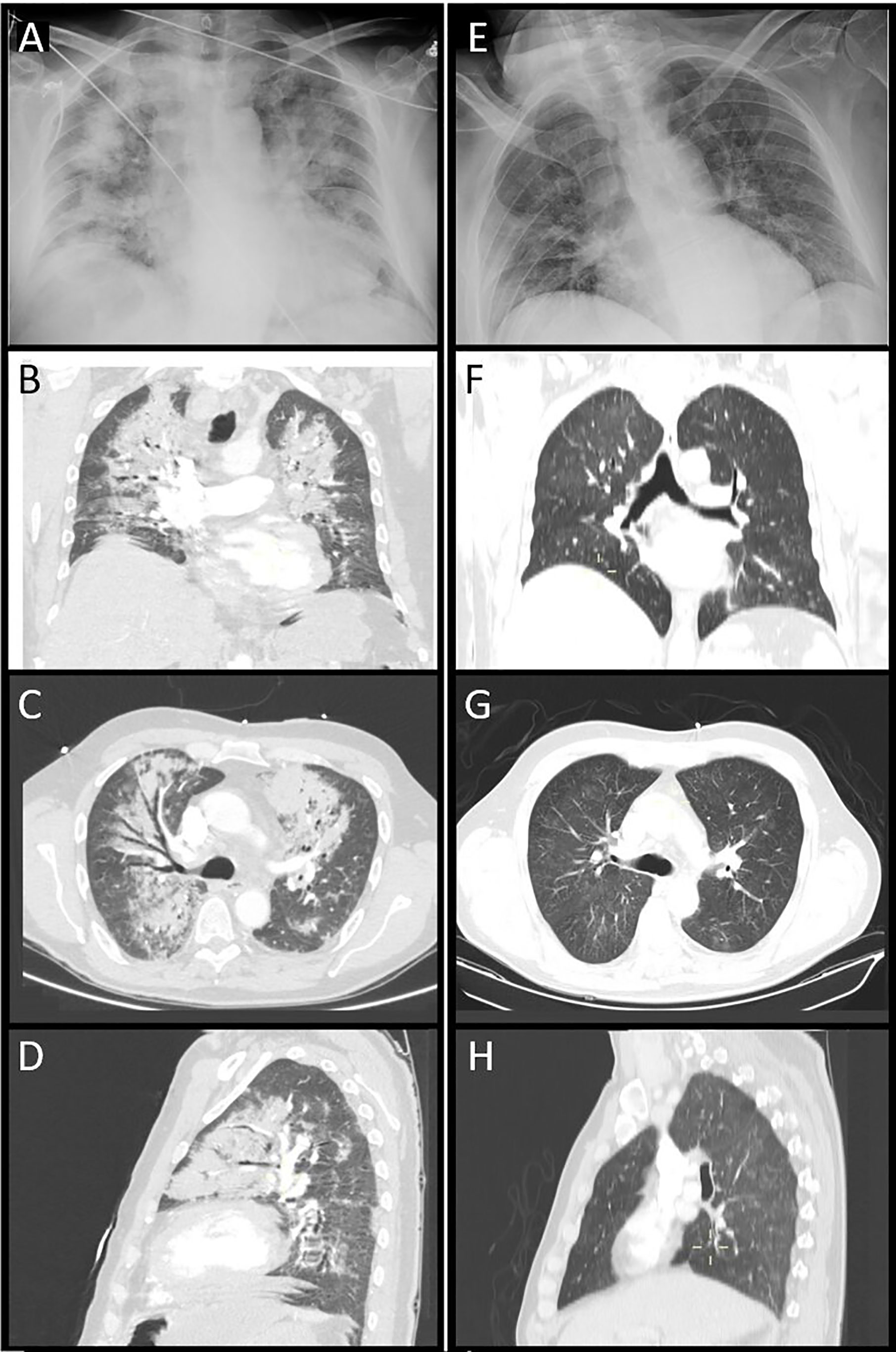
Chest X-ray and computed tomography on admission and the 28th day after ...
Paradoxical respiration: ‘Seesaw’ motion with massive pulmonary ...
Study Finds COVID-19 Less Seve [IMAGE] | EurekAlert! Science News Releases
Clinical management of lung cancer patients with respiratory symptoms ...
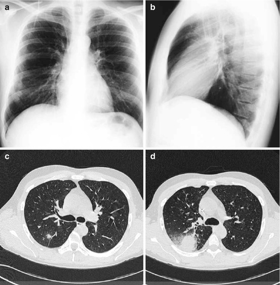
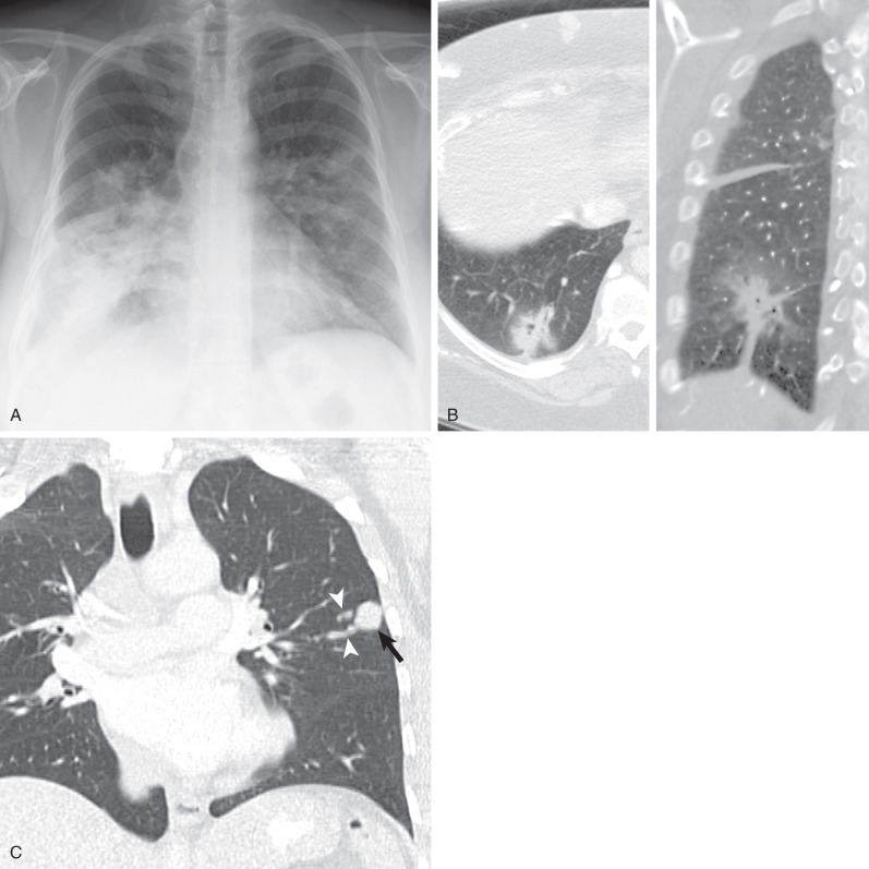
Chest radiography and CT images of a 30-year-old woman with cough and ...
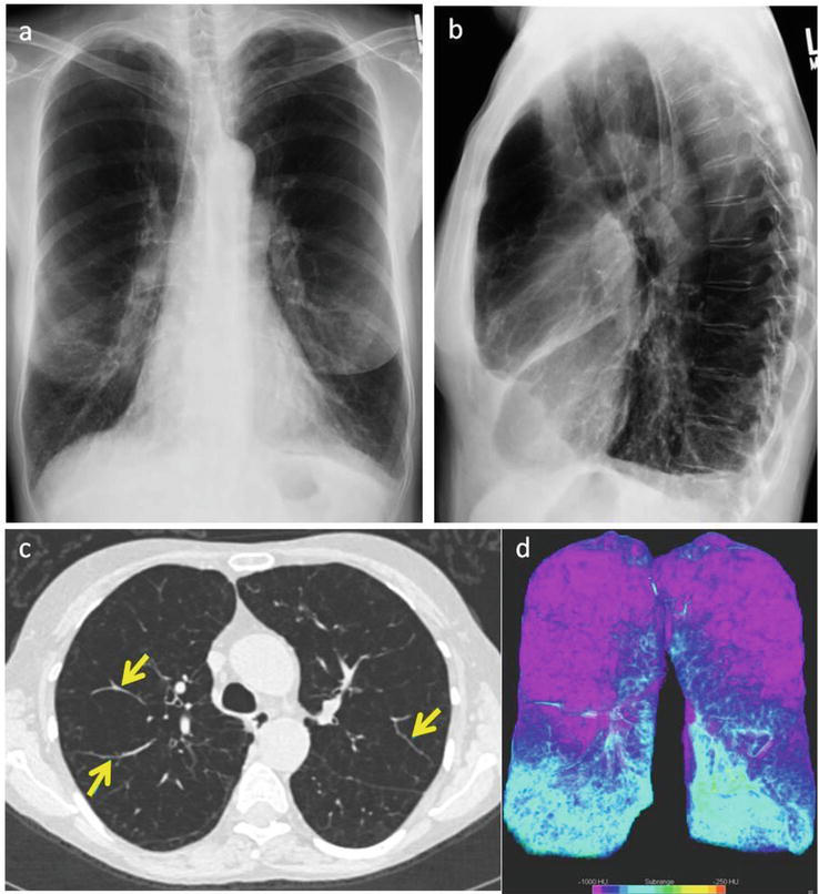
Pulmonary fibrosis on the lateral chest radiograph: Kerley D lines ...
Whole lung lavage in alveolar proteinosis: manual clapping versus ...
Imaging of Emphysema: A Comprehensive Review | IntechOpen
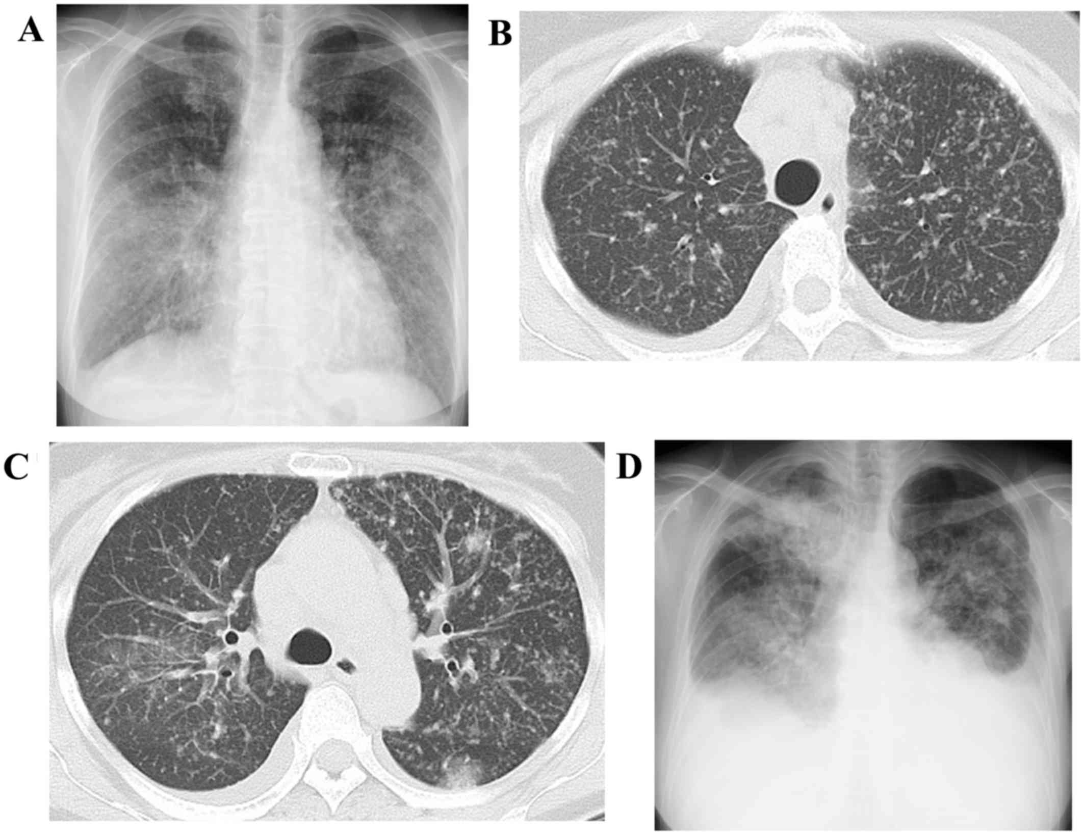
(A) Chest X-ray showing a micronodular pattern. (B) HRCT scan showing ...
Synchronous neuroendocrine tumor and non‐small‐cell lung cancer in ...
Diagnostic Radiology in Hematological Patients with Febrile Neutropenia ...
Micronodular pattern of organizing pneumonia: Case report an... : Medicine
Fungal Infections | Radiology Key
Frontiers | Catecholaminergic Crisis After a Bleeding Complication of ...
The pulmonary nodule following lung transplantation - Clinical Imaging
| the spectrum of Ra-associated lung disease. a | A CT image of a ...
Miliary lung metastases from non‑small cell lung cancer with Exon 20 ...
Case 20-2014 — A 65-Year-Old Man with Dyspnea and Progressively ...
Pathophysiological mechanism of immersion pulmonary edema onset in this ...
Figure2.Representative clinical images of Eisenmenger syndrome. A ...
Update on diffuse alveolar hemorrhage and pulmonary vasculitis ...
Diagnostic utility of transbronchial biopsy for Hodgkin's lymphoma: A ...
a, b Chest X-ray of endobronchial coils without signs of infection. c ...
Imaging Primer on Chimeric Antigen Receptor T-Cell Therapy for ...
Intralobular septal thickening on chest CT in a patient with pulmonary ...
7 Differences Between X Ray And CT Scan - Deep Medical Centre
Figure1.(a, b) On plain chest radiography, bilateral alveolar opacities ...
A 6-year-old boy with lymphoma (patient 4) and fever. He had ...
Synchronous Pneumatocele and Organizing Pneumonia During Staphylococcus ...
Chest X-ray showed infiltrates in the bilateral lungs and right pleural ...
Thoracic radiography showed infiltrates in the entire right lung field ...
Figure1.a and b: Chest x-ray and computed tomography scan showing ...
Miliary Nodularity in a Patient Receiving TNF Inhibitors Is Not Always ...
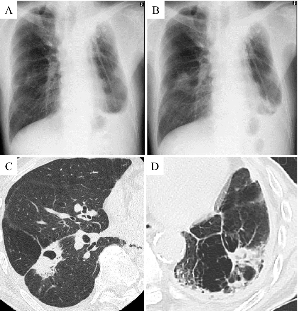
Chest radiography and computed tomography. (A) Chest radiograph before ...
Figure9.Chest radiograph (A) and high-resolution chest CT scan (lung ...