Please enter url.
Login
Logout
Please enter url.
Ct Scan Enlarged Lymph Nodes
mavink.com
source
Comments
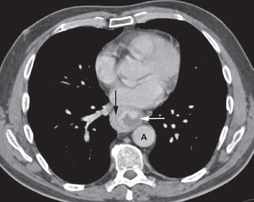
Figure 3:Burkitt lymphoma masquerading as cardiac tamponade- Open-i
Video-Assisted Thoracic Surgery for Diseases Within the Mediastinum ...
Gastrointestinal Tract | Radiology Key
Intraoperative finding of an infected composite graft (arrow ...
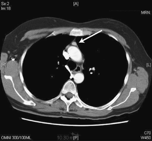
CT Scan showing a tumorous lesion affecting the upper anterior ...
The chest X-ray and chest CT show an endobronchial mass in the left ...
Figure 1 from Acute Chest Pain Related to Pericardial Fat Necrosis ...
The white arrow is pointing to the right internal mammary artery ...
Chest radiograph appearances of an angiolipoma in a 71 year old female ...
Azygos vein aneurysm - A rare entity | Eurorad
Mediastinal lymph node postoperative pathology (H & E staining, ×200 ...
Pulmonary LELC with local recurrence in right lung (pre-resection ...
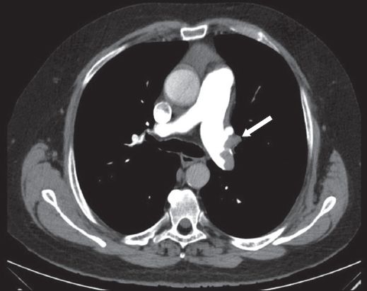
CT scan of the thorax demonstrating necrotic paratracheal node ...
Cardiothoracic ratio was calculated from axial CT images by dividing ...
Internet Scientific Publications
Axillary lymphadenopathy on CT thorax with contrast. | Download ...
Chest computed tomography (CT) scan shows a right paratracheal cystic ...
CT demonstration of vertical veins | Eurorad
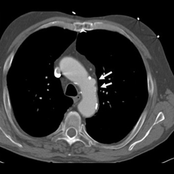
Fusiform ascending aortic aneurysm—sagittal reconstructed CT image in a ...
Aortic ductus diverticulum mimicking aortic dissection: a case of ...
Coronal and axial neck and chest CT scan shows asymmetric nodular ...
Masaoka staging system for thymoma | Download Table
Axial CT image demonstrates pulmonary embolism within the left ...
Enlarged Mammary Glands
Chest CT revealing a pulmonary nodule (white arrows) before surgery ...
Left superior vena cava as an incidental finding in a 70-year-old man ...
CT chest without contrast showing a new 2.2 x 1.7 x 4.5 cm soft tissue ...
Enhanced CT showing an enlarged axillary lymph node (white arrow ...
Brucella melitensis sternal osteomyelitis following median sternotomy ...
Chest X-ray showing left basal pneumonia with para-pneumonic effusion ...
Electrocardiographic findings in pulmonary embolism | SMJ
Figures
Endoscopic views of the patient. a Downhill varices in the upper third ...
(PDF) Large Retrosternal Goiter: An Otolaryngological Perspective ...
(PDF) Aortitis as a cause of severe abdominal pain
Aortic-Lymph-Nodes-Cancer
Aortic-Arch-Lymph-Nodes
Para-Aortic-Nodes
Left-Para-Aortic-Lymph-Node
What-Are-Para-Aortic-Lymph-Nodes
Peri-Aortic-Lymph-Node
Para-Aortic-Lymph-Nodes-Location
Enlarged-Lymph-Nodes-Ultrasound
Iliac-Lymph-Nodes-Location
Lymph-Nodes-Aorta
Para-Aortic-Lymph-Node-Dissection
Hilar-Lymph-Nodes-Lung-Cancer
Celiac-Lymph-Nodes-Location
Aortocaval-Lymph-Node
CT-Abdomen-Lymph-Nodes
Enlarged-Lymph-Nodes-Near-the-Groin