Please enter url.
Login
Logout
Please enter url.
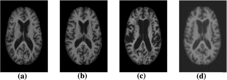
Shown are the (a) MPRAGE, (b) FLAIR, (c) T 2 -w, and (d) PD-w images ...
researchgate.net
source
Comments
Brain MRI analysis for Alzheimer’s disease diagnosis using an ensemble ...
Transient Splenial Lesion of the Corpus Callosum in Clinically Mild ...
(a) Rician distribution of y (Sec. 2.1) for different values of v ...
Two-dimensional grid image based on brain MRI images brightness and ...
Clinics in diagnostic imaging (193) | SMJ
Axial section of IBSR data: the original image (a), the segmented brain ...
Sample images from OASIS database. (a) Sample category-1 MRI image, (b ...
Data acquired under different pulse sequences or different scanners ...
Residual inception (RI) block for tumor extractor | Download Scientific ...
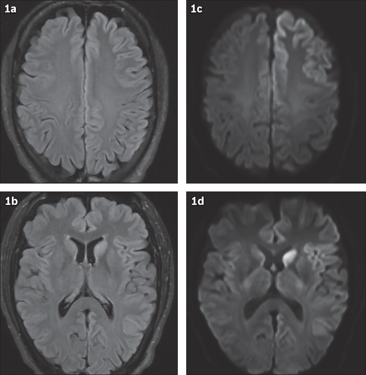
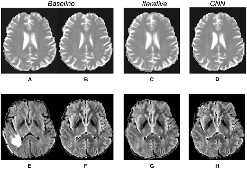
Diffusion-Weighted Image and Diffusion Tensor Imaging
Conclusion Diffusion at high b values on clinical scanner was shown to ...
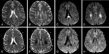
Example images of the motion categories used for statistics: invisible ...
Diffusion-Weighted Magnetic Resonance Imaging in Acute Stroke | Stroke
Is the “biexponential diffusion” biexponential? - Kiselev - 2007 ...
(1) Patient with no dementia; (2) patient with very mild AD; (3 ...
A: axial FLAIR image shows multifocal cortical and subcortical ...
Experiment on simulated image: (a) Original Image (simulated with 3% ...
Diagnostics | Free Full-Text | HTLML: Hybrid AI Based Model for ...
Frontiers | Quantitative evaluation of the influence of multiple MRI ...
(a) FLAIR (b) T1-w (c) T1-w contrast enhanced (d) T2-w MRI brain Images ...
Four axially reformatted images from an MS patient, no contrast ...
DWI differentiates MSA-P from PSP, Parkinson's disease and controls ...
| Diffusion-weighted images of the study sample. (A) Patient No. 1; (B ...
A sample slice from the BrainWeb: (a-c) Input slice: T1, T2 and PD ...
Patient 3. MR imaging findings at the time of neurologic event (A-C ...
The figure shows the effect of N4 bias correction on a FLAIR image with ...
The MRI sequences vs. the standardized image. Images (a) and (b) show ...
Shows comparison of Normal and Demented MRIs [2] (a) no dementia, (b ...
Accuracy and reproducibility of manual and semiautomated quantification ...
Association of MRI Markers of Vascular Brain Injury With Incident ...
Figure 1 from Automatic detection and classification of Alzheimer's ...
Four different MRI pulse sequences of brain tumor | Download Scientific ...
Brain parenchymal regions in a T2-weighted image, b T1-weighted image ...
Randomly samples deformation fields can yield anatomically implausible ...
28 years-old Female patient with RRMS (4 years duration) and normal ...


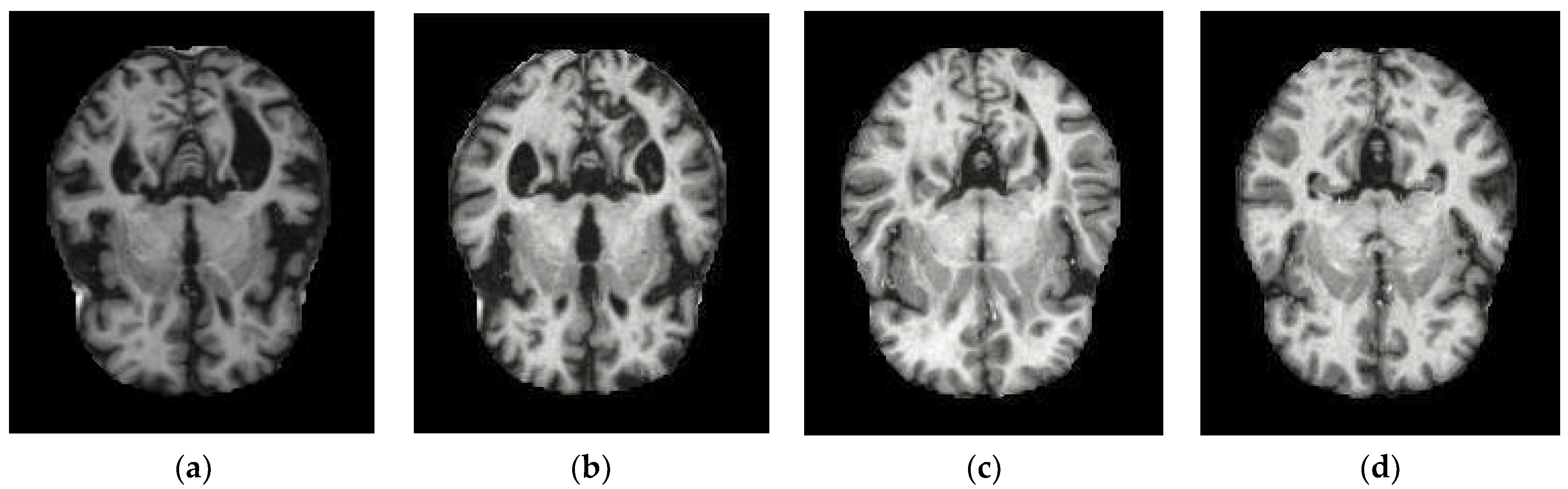


![Shows comparison of Normal and Demented MRIs [2] (a) no dementia, (b ...](https://www.researchgate.net/profile/Gururaj-Awate-2/publication/330727294/figure/fig1/AS:720653959319554@1548828701448/Shows-comparison-of-Normal-and-Demented-MRIs-2-a-no-dementia-b-very-mild-dementia_Q640.jpg)


