Please enter url.
Login
Logout
Please enter url.
Normal Female Ct Scan Of Abdomen With Labels
mavink.com
source
Comments
(a) Dilated intrahepatic bile ducts observed on abdominal CT (black ...
Diagnostics | Free Full-Text | Prognostic Assessment of ...
Medical Treatment of a Staghorn Calculus: The Ultimate Noninvasive ...
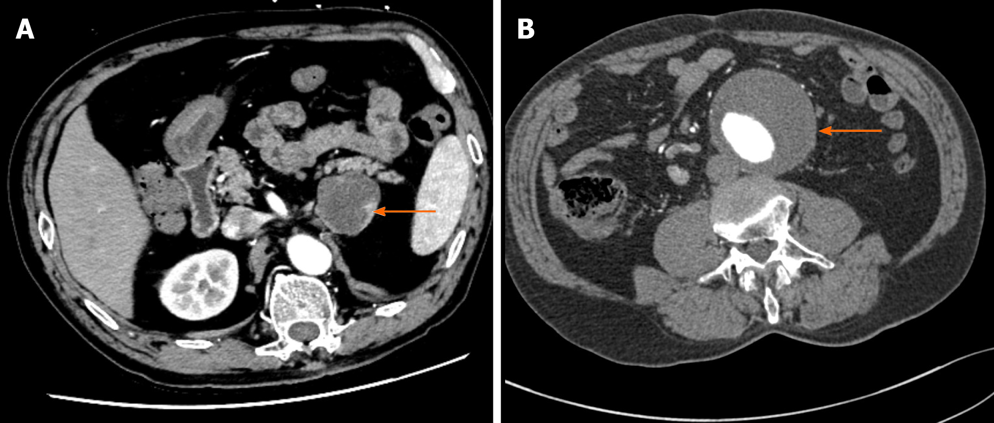
Pheochromocytoma with abdominal aortic aneurysm presenting as recurrent ...
Cureus | Primary Renal Carcinoid: Two Rare Cases at a Single Center
A literature review of radiological findings to guide the diagnosis of ...
Frontiers | Central and Extrapontine Myelinolysis Affecting the Brain ...
Cross sectional images of contrast enhanced computerized tomography ...
a) Axial contrasted CT scan showing dilated small bowel loops with ...
Adrenal Cushing syndrome with detectable ACTH from an unexpected source ...
Upper gastrointestinal bleeding: gallstone-induced auto-sphincterotomy ...
Adult duodenal intussusception associated with congenital malrotation
Abdominal and pelvic computed tomography scan. A, B: The computed ...
Visceral Arterial Compromise During Intra-Aortic Balloon ...
A case of primary malignant fibrous histiocytoma of the pancreas: CT ...
(PDF) Correlation between modified CT severity index and ...
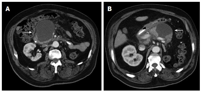
A large series of true pancreaticoduodenal artery aneurysms - Journal ...
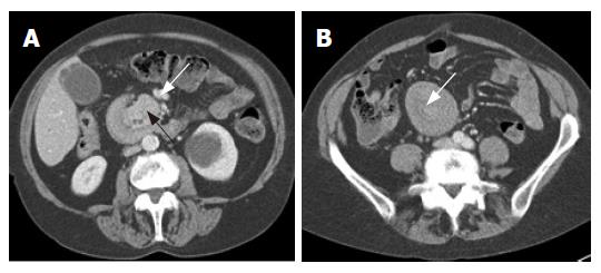
(A) CT shows two limbs of the fluid-filled afferent loop (arrows ...
Axial CT scans of the abdomen. Duodenal GIST described | Open-i
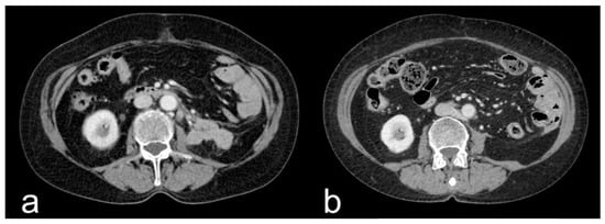
Contrast-enhanced CT 6 months ago found suspicious nodules at the ...
Computed tomography of the abdomen with contrast. (a) A large ...
Applied Sciences | Free Full-Text | Advanced CT Imaging, Radiomics, and ...
An atypical presentation of intrahepatic perforated cholecystitis: a ...
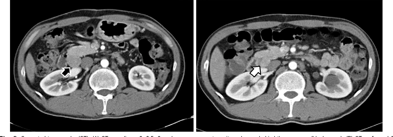
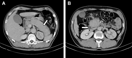
Retroperitoneal haemorrhage secondary to spontaneous rupture of giant ...
Medical Treatment of a Staghorn Calculus: The Ultimate Noninvasive ...
State of the Art of CT Detectors and Sources: A Literature Review ...
Transverse section of abdominal CT scan. There is a marked abnormality ...
A: Noncontrast axial CT of the abdomen showing increased volume of the ...
Dual phase abdominal contrast CT scan. Tumour at the bifurcation of the ...
Figure 1 from Synchronous Peripancreatic Lymph Node Gastrinoma and ...
Figure 14:The Utility of 64 Channel Multidetector CT Angiography for ...
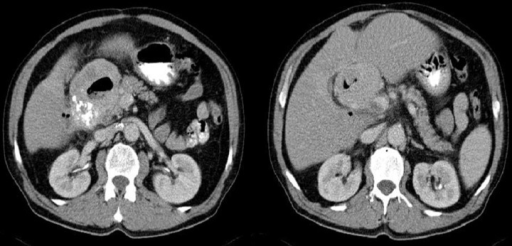
Immunoglobulin G4-related disease presenting with obstructive jaundice ...
Radiology of pancreatic neoplasms: An update
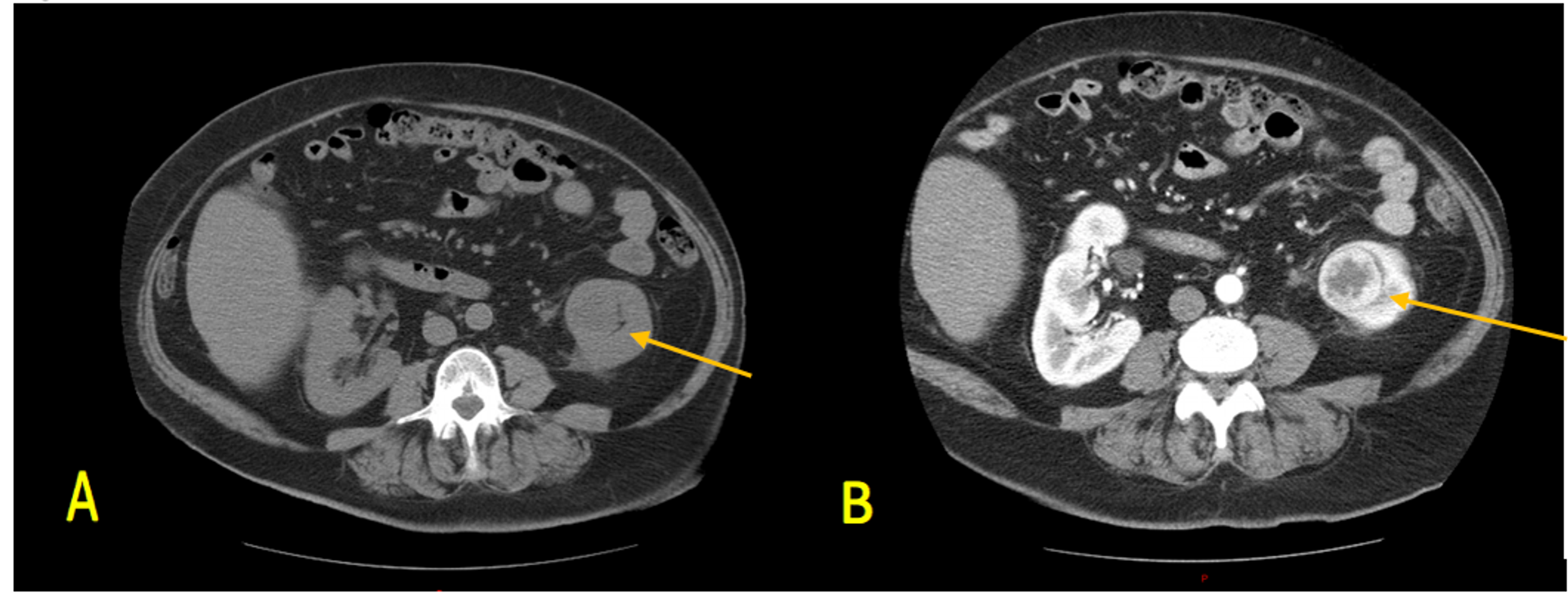
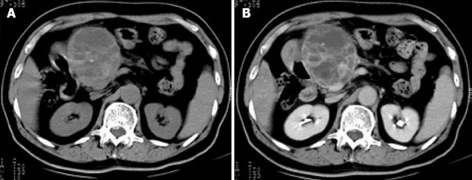
Solitary pancreatic renal cell carcinoma metastasis
Initial diagnostic abdominal CT scan. No abnormal findings of superior ...
Abdominal-CT-Scan-Colon-Cancer
Cat-Scan-Abdomen
Abdominal-CT-Scan-Labeled
Liver-CT-Scan
Abdominal-Hernia-CT-Scan
Abnormal-CT-Scan-Abdomen
Abdominal-and-Pelvic-CT-Scan
Lymphoma-CT-Scan
Stomach-CT-Scan
Abdominal-CT-Scan-without-Contrast
CT-Scan-Test
Male-Pelvis-CT-Scan
Normal-CT-of-Abdomen
Pancreas-CT-Scan-with-Contrast
Abdominal-MRI-Scan
Chest-CT-Scan