Please enter url.
Login
Logout
Please enter url.
Magnetic resonance imaging brain. (a-c) T2-weighted axial section ...
researchgate.net
source
Comments
The axial Trace (a) and ADC (b) DWI images shows restricted diffusion ...
MR Imaging Can Predict the Development of Nonalcoholic Wernicke ...
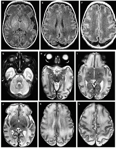
Axial magnetic resonance images in a 3-month-old girl to evaluate ...
Differential diagnosis of infarct‐like intracranial ectopic germinomas ...
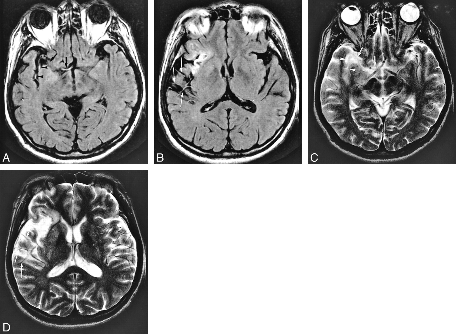
MRI of the brain, FLAIR sequence (TR/TE/FA: 7000/135/150). (A-C): Axial ...
Transverse T1-weighted magnetic resonance images of the brain. Notes ...
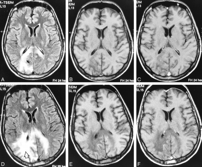
Effect of MR sequence on visibility of the pulvinar sign. A ...
MRI of Patient 2, revealing multiple lacunar infarctions of frontal ...
Bilateral Rasmussen encephalitis - Epilepsy & Behavior
Case 27-2018: A 3-Year-Old Boy with Seizures | New England Journal of ...
Intraventricular Hemorrhage - Cytotoxic Edema - Mussen Healthcare
Imaging findings of patients with MBD. Transverse diffusion-weighted ...
Early-onset Combined Methylmalonic Aciduria and Homocystinuria ...
Cerebral Microbleeds: Incidence, Imaging Characteristics, Common and ...
Coinfection of Japanese Encephalitis with Neurocysticercosis: An ...
Figure 1 from Myotonic Dystrophy Type 1 Associated with White Matter ...
Fluid-attenuated Inversion Recovery Intraarterial Signal: An Early Sign ...
Highly Active Antiretroviral Therapy for Patients with AIDS Dementia ...
Disseminated Aspergillosis Involving the Brain: Distribution and ...
Normal Development of the Fetal, Neonatal, and Infant Brain, Skull, and ...
Figure 1 from Diffusion-weighted and gradient echo magnetic resonance ...
Patient 1. Images at presentation (A) and follow-up 5 (B-E) and 45 (F ...
Normal Development of the Fetal, Neonatal, and Infant Brain, Skull, and ...
Top 10 Rarest Diseases Can Infect The Human | TopTeny.com
Silent Brain Infarcts | Stroke
Magnetic Resonance Imaging - Magnetic Resonance - 78 Steps Health
Identifying Subtle Cortical Gyral Abnormalities as a Predictor of Focal ...
Initial MRI taken on admission. (A,B,C,D) FLAIR images show multiple ...
Diagnosing Variant Creutzfeldt-Jakob Disease with the Pulvinar Sign: MR ...
A 5-year-old girl with left cerebellar JPA. Residual tumor failed to ...
Focal neuronal loss, reversible subcortical focal T2 hypointensity in ...
Case 1. A 40-year-old woman who presented with fever, chill and altered ...
MRI in CLN2 disease patients: Subtle features that support an early ...
Central nervous system anomalies on MRI T2-weighed images in two ...
Differential diagnosis of infarct‐like intracranial ectopic germinomas ...