Please enter url.
Login
Logout
Please enter url.
Ultrastructure of the liver hepatocyte. Electron micrograph: H ...
researchgate.net
source
Comments
Ultrastructure of the liver hepatocyte. Electron micrograph: H ...
MyoWbroblast in the cortical peritubular interstitium in a rat kidney ...
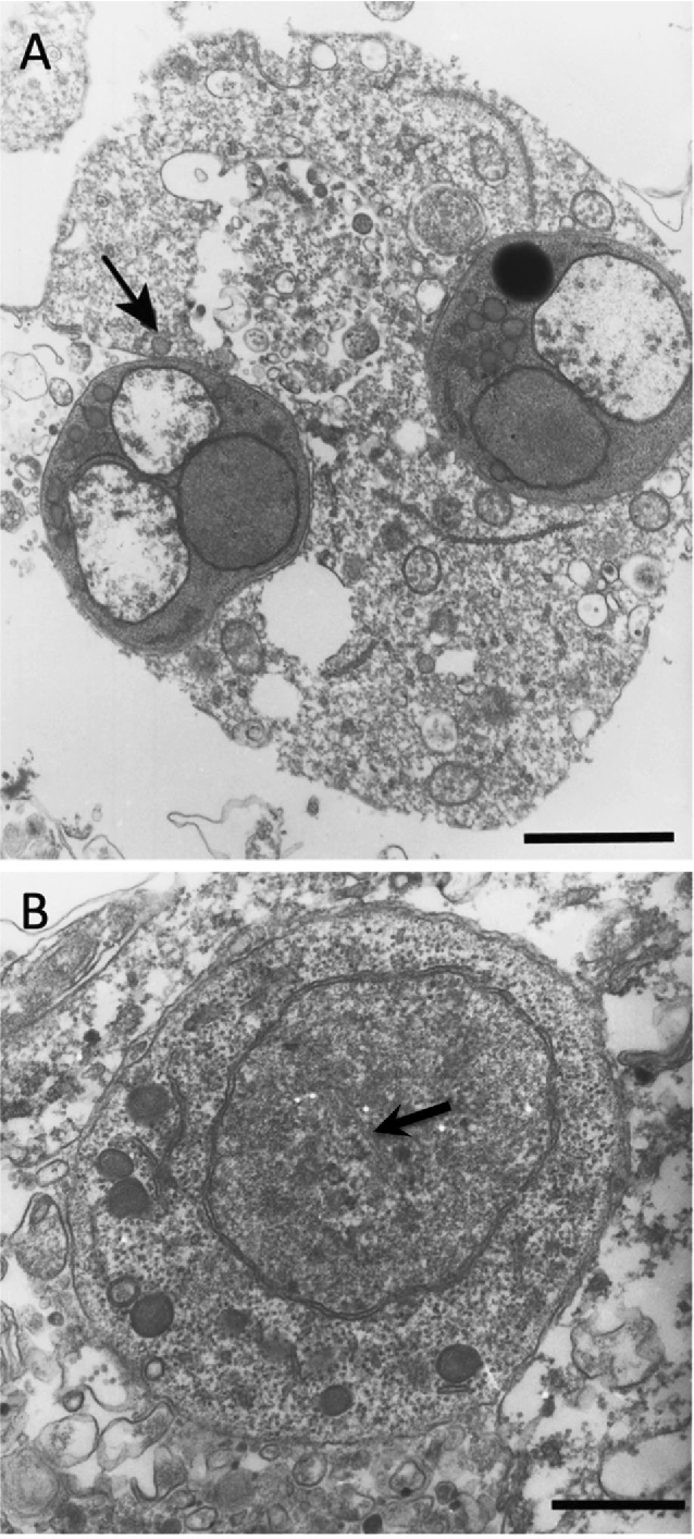
Memórias do Instituto Oswaldo Cruz - Interaction and cystogenesis of ...
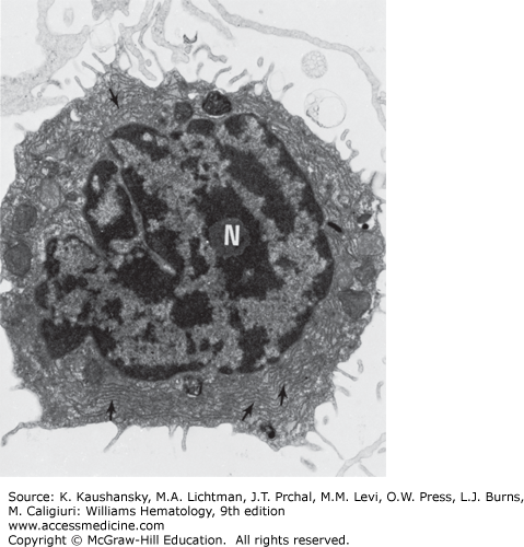
The Structure of Lymphocytes and Plasma Cells | Oncohema Key
Electron microscopy analysis of four cell types infected with VTT ...
Figure 3. | Establishment and Characterization of Three Novel Cell ...
A Road Down Memory Lane: Biology... Exploring Levels of Biological ...
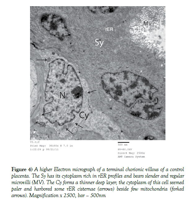
Study of the Ultrastructure of the placenta in gestational Diabetes ...
Changes in cytoplasm organization in degenerating early embryos of D ...
Pyriform cell differentiation in Podarcis sicula is accompanied by the ...
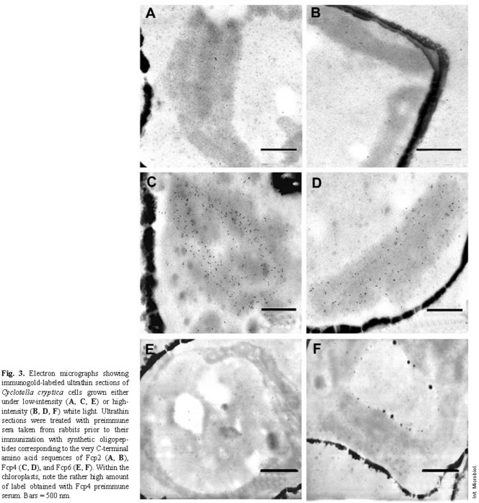
Cuantificación por microscopia inmunoelectrónica de los polipéptidos ...
SciELO - Brasil - Functional cytology of the hepatopancreas of ...
Ultrastructural Changes of Neuronal Mitochondria After Transient and ...
SciELO - Brasil - Ultrastructural alterations of choroid plexuses of ...
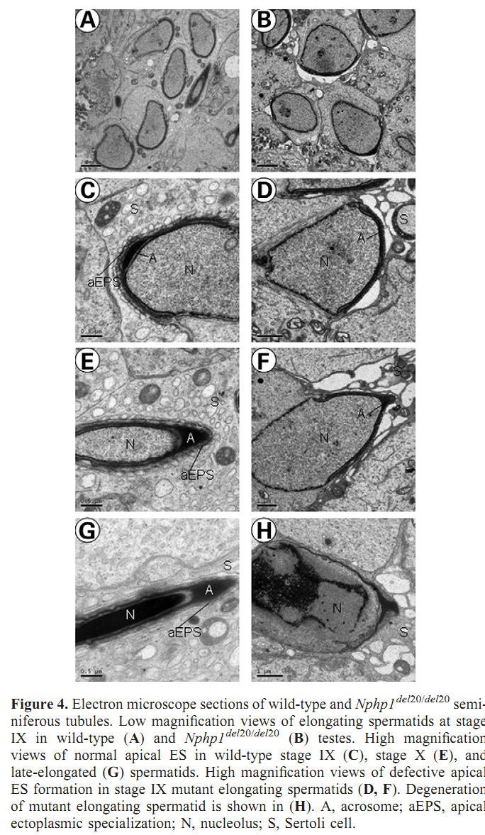
FertilityOnline | Spermatogenes
Full article: Pharmacological, toxicological and neuronal localization ...
Formation of Mallory Body-like Inclusions and Cell Death Induced by ...
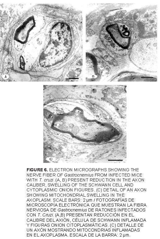
Gastrocnemius skeletal muscle microvasculature and neuromuscular ...
(a) NRK-52E cells were imaged using SBF-SEM at 3,500x, with a voxel ...
Sample FAb 74, GW2 section, see Fig. 12 for sample location ...
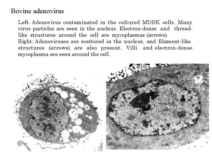
NIAH:NIAH Pathogenic Organisms Observed by Electron Microscope:Adenovirus
Mitochondrial alterations in diabetes. (a) Normal (arrows) and swollen ...
Structure of posterior semicircular duct | Semantic Scholar
Figure 2 from Ultrastructural comparison of Bonamia spp. (Haplosporidia ...
The Golgi complex of epididymal principal cells. A-C. Transmission ...
Transmission electron micrograph of the vaginal epithelium during the ...
The TEM analysis of ultrastructural mitochondrial changes after ...
Electron micrograph of the lung of calf no 3 that died 36 hours p.i ...
Figure 1 from TEM observation of ultrasound-induced mitophagy in ...
Transmission electron micrograph, splenic neoplasm, dolphin 5 ...
Unusual cytoskeletal structures found in the cytoplasm of cryoWxed ...
Electron micrograph. Cecum; C3H/HeOuJ mouse 6 hours following infection ...
A Comparative Study of Tunica Media of Some Arteries in Postnatal Rats ...
Latex beads coated with Mce1D was internalized by HeLa cells. (a) Latex ...