Please enter url.
Login
Logout
Please enter url.
EPOS™
epos.myesr.org
source
Comments
3D organization and images of vertical sections of the gills from a ...
DR5:GUS Expression and Root Development in sfc Mutants. | Download ...
First description of epimorphic development in Antarctic Pallenopsidae ...
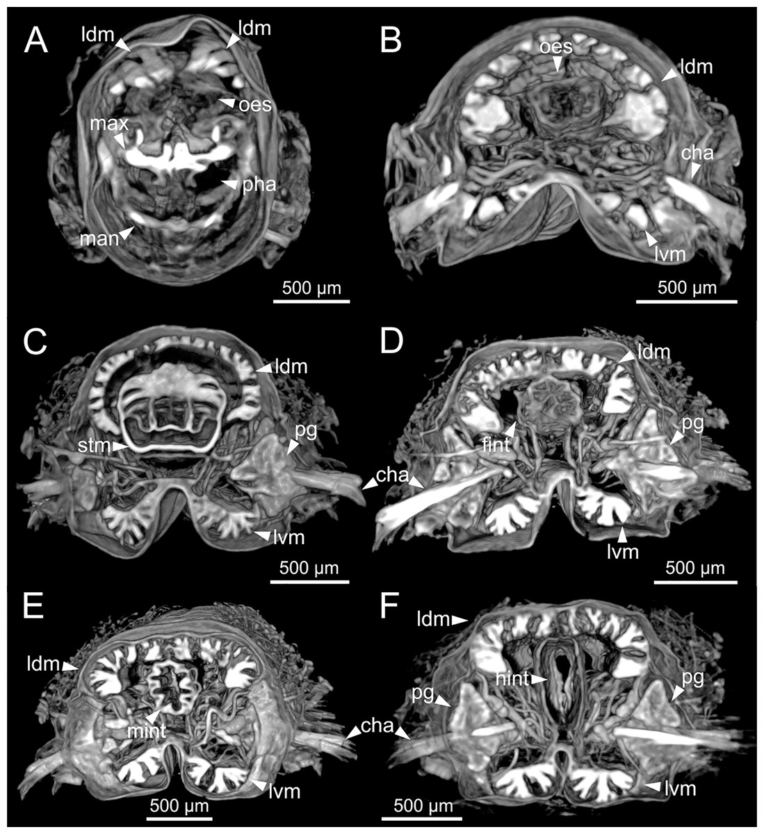
A quantitative micro-tomographic gut atlas of the lepidopteran model ...
Surface representation of RECK-His. A, three-dimensional reconstruction ...
Images of Shevtchenkella caboata sp. nov. (A, B and D) Prodorsal ...
Nodoprosopidae new family. 1, Nodoprosopon ornatum , NHMW... | Download ...
Figure 2 from Visualization of microvasculature by x-ray in-line phase ...
3-D depiction of likely damage in Phineas Gage (Panel A) [Reprinted ...
Practical Illustrations Sodium Spectroscopy and Imaging: Clinical ...
-figure supplement 1: Cribriform plate morphology in Swiss Webster and ...
Results obtained in the classification step for two 3D images. (a) One ...
ASN1‐encoded asparagine synthetase in floral organs contributes to ...
Material Decomposition - Spectral, Photon Counting Computed Tomography ...
[PDF] Tensor completion and low-n-rank tensor recovery via convex ...
Konetontli migrus, n. sp., habitus, holotype-(CNAN T0891). A. Dorsal ...
Vascular Precursors and Ground Tissue Stem Cells Divide Abnormally in ...
Reducing distortions in diffusion‐weighted echo planar imaging with a ...
Larval and Pupal wings of Bicyclus anynana. (A) Early larval forewing ...
The development of primary HCCs in rats treated with intraperitoneal ...
Applied Sciences | Free Full-Text | 2D/3D Multimode Medical Image ...
Laser scanning confocal images of tick synganglia showing 5HT-IR in ...
(PDF) Endoscopic Trans-Lateral Molar Approach to Infratemporal Fossa ...
Reconstructed optoacoustic image using non-negative constrained model ...
Ovule development revealed by confocal laser scanning microscopy in ...
MIP is expressed in apical organ neurons in Platynereis and Capitella ...
Long forgotten: Eunice woodwardi Baird, 1869 (Annelida, Eunicidae ...
Brain nuclei receiving Brn3c Cre/WT ; ROSA26 tdTomato/WT neuronal ...
(Top) Typical examples of images obtained from low-dose CT, water data ...
Wydundra leichhardti sp. nov., male (A–F) and female (G-H). A ...
Qualitative denoising results on the Brain-Tumor-Progression [12] MRI ...
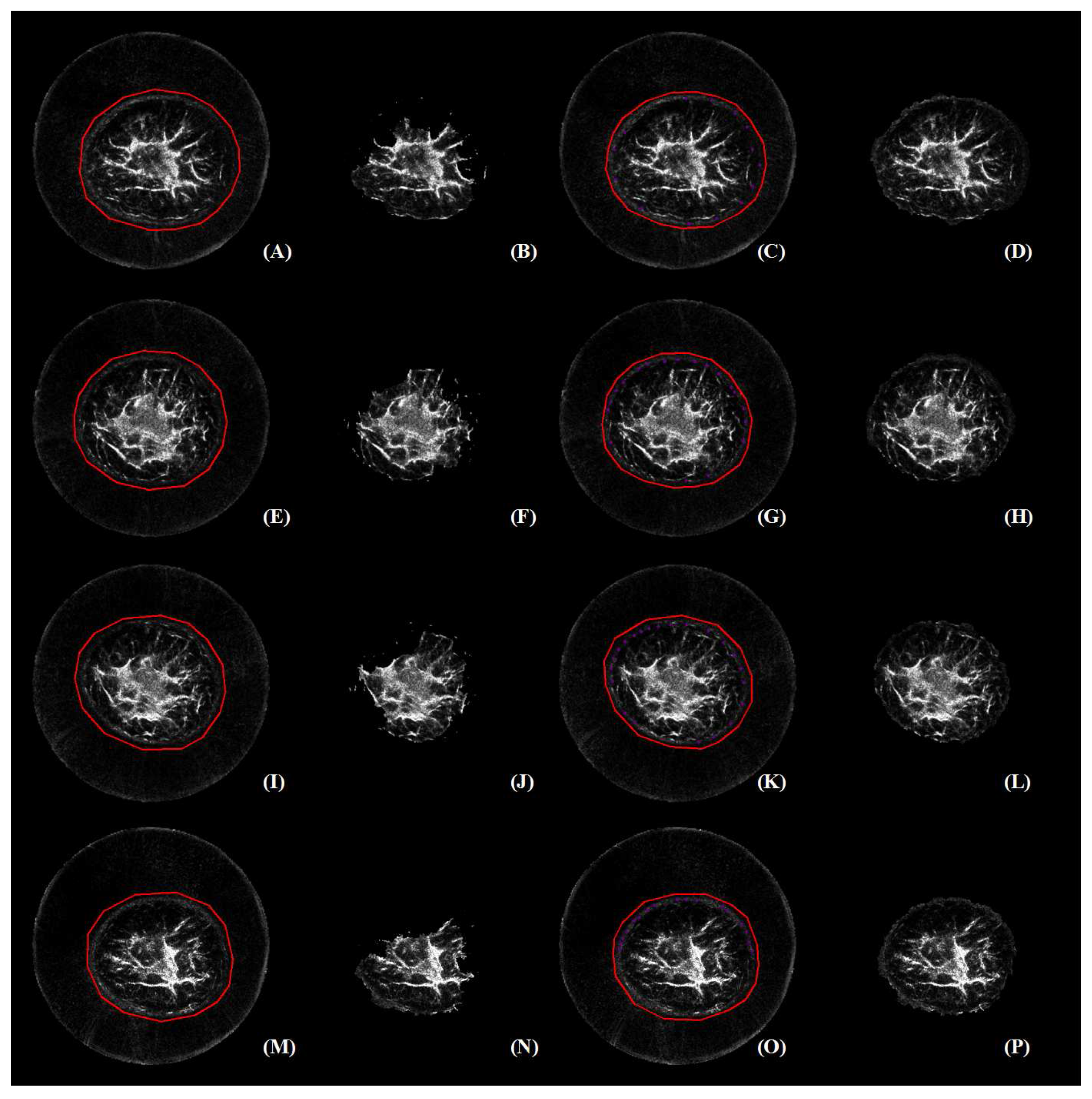
Evaluation of fibroblasts migration and wound closure using ...
Sensors | Free Full-Text | Efficient Segmentation of a Breast in B-Mode ...
Identification of Neural Networks That Contribute to Motion Sickness ...
Representative multimodality QT ultrasound images of a volunteer's ...


![[PDF] Tensor completion and low-n-rank tensor recovery via convex ...](https://d3i71xaburhd42.cloudfront.net/f59019768cc6e198b73d52adcfaa0d26067a6e28/500px/17-Figure3-1.png)


![Qualitative denoising results on the Brain-Tumor-Progression [12] MRI ...](https://www.researchgate.net/publication/366190352/figure/fig2/AS:11431281106626237@1670814988673/Qualitative-denoising-results-on-the-Brain-Tumor-Progression-12-MRI-test-data-set-of-T1_Q640.jpg)