Please enter url.
Login
Logout
Please enter url.
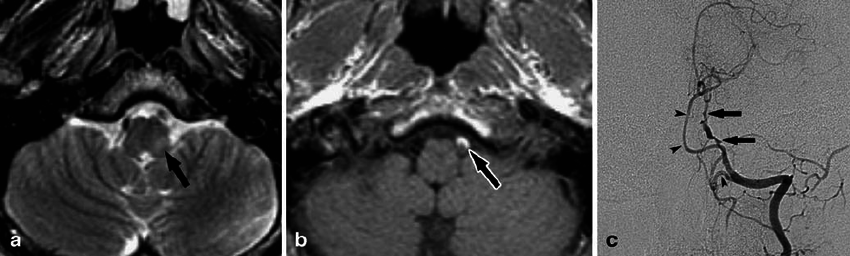
Figure 4 from Subarachnoid Hemorrhage Confirmed by Magnetic Resonance ...
semanticscholar.org
source
Comments
BIR Publications
Initial pretreatment images. (a and b) Magnetic resonance imaging ...
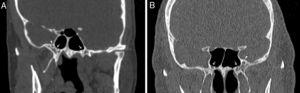
Endolymphatic sac tumor. a Axial CT image shows an expansile ...
Images from a 38-year-old man with left-sided facial pain, vertigo, and ...
FH (white arrow) in the sagittal image (A), in the axial image (B ...
Non-pulmonary Artery Thrombus | Radiology Key
Magnetic resonance vessel wall imaging in cerebrovascular diseases in ...
Ramsay Hunt Syndrome Associated with Brain Stem Enhancement | American ...
Persistent primitive hypoglossal artery variant. Right external carotid ...
Norethisterone enanthate-induced cerebral venous sinus thrombosis (CVST ...
Time-of-flight imaging in a 4-year-old boy with occipital horn syndrome ...
Trauma | Radiology Key
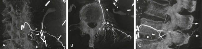
Arterial Anatomy of the Spine and Spinal Cord | Radiology Key
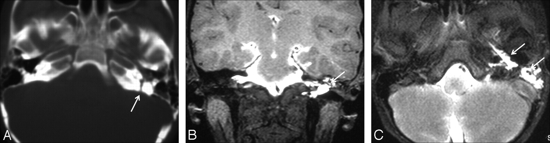
The anatomic analysis of the vidian canal and the surrounding ...
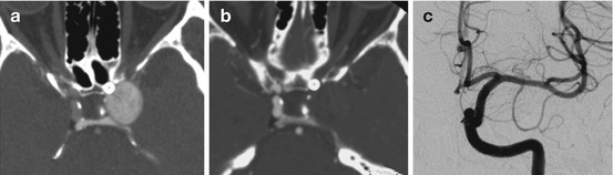
Preoperative magnetic resonance images. (a) A vascular lesion is ...
Delayed neurological deterioration following atlantoaxial facet joint ...
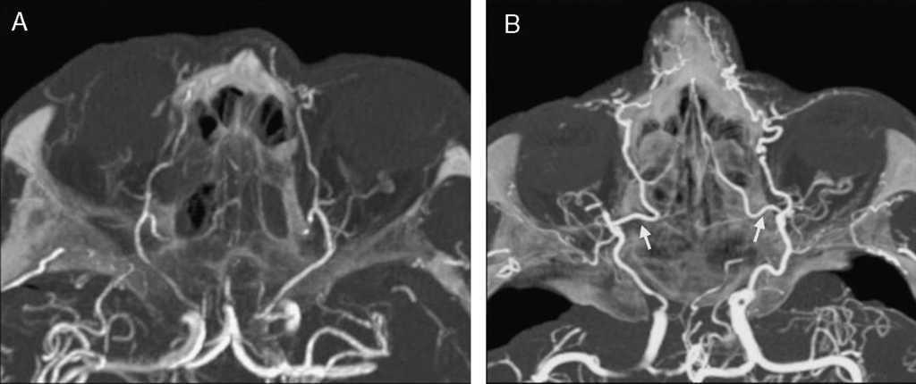
Radiological Anatomy of the Ethmoidal Arteries: CT Cadaver Study | Acta ...
Neurovascular Imaging at 1.5 Tesla Versus 3.0 Tesla - Magnetic ...
Intrathecal Gadolinium-Enhanced MR Cisternography in the Evaluation of ...
Stents in the Treatment of Intracranial Aneurysms | Radiology Key
Dysgenesis of the Internal Carotid ArteryAssociated with ...
MR Venography in the Pediatric Patient | American Journal of Neuroradiology
Lateral Medullary Syndrome and Ipsilateral Hemiplegia (Opalski Syndrome ...
Temporomandibular joint scalograms. A: Closed mouth vie | Open-i
Imaging Manifestations and Interventional Treatments for Hereditary ...
Inflammatory Pseudotumor of the Nasopharynx and Skull Base: Mimicking ...
Chordoma and Chondrosarcoma | Neupsy Key
A, Axial MIP image of a term-born infant in which the AcomA cannot be ...
CT and MR Imaging of Giant Cell Granuloma of the Craniofacial Bones ...
Early postoperative MRI and detection of residual adenoma after ...
male patient, 70 years. Axial Time-of-Flight (TOF) and 3D... | Download ...
Essentials for Interpreting Intracranial Vessel Wall MRI Results: State ...
Controversies in the Management of Geriatric Odontoid Fractu ...
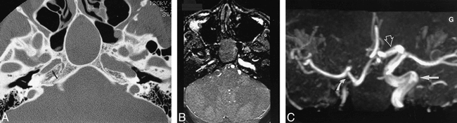
A , CTA, transversal MIP reconstruction; image thickness 10 mm. B ...
Simple DNT variant in the right amygdala. Sagittal T1weighted 3D-MDEFT ...