Please enter url.
Login
Logout
Please enter url.
Patent Ductus Arteriosus Echocardiography
mavink.com
source
Comments
Fibroepithelial polyp arising from the posterior vaginal wall ...
(PDF) Mitral degenerative disease mimicking a valvular tumor: A case report
Pre-procedural echocardiographic images. (A) Apical 4-chamber view ...
TEE four-chamber view showing chordal SAM (arrows) (RV: Right ...
Echocardiographic atypical parasternal long axis view of the tricuspid ...
CT angiogram done after bidirectional Glenn operation showing good ...
Transesophageal echocardiography (TEE). Perivalvular cavity around the ...
Midesophageal aortic valve long-axis view showing presence of a ...
Continuous-wave Doppler tracing through the tricuspid valve. | Download ...
After evacuation of the hematoma, mid-esophageal colorflow Doppler ...
Echo Mv Area By Pressure Half Time - Echocardiography
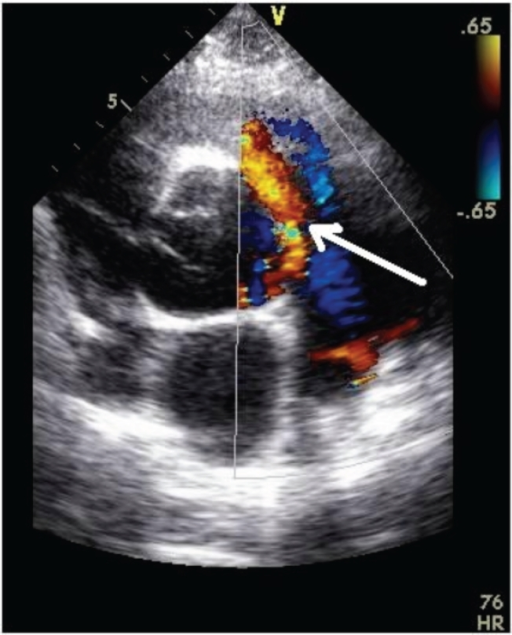
Echocardiogram demonstrating juxtaductal coarctation of the aorta ...
Four-chamber echocardiography view of PV stenosis in patient with FM ...
Figure. Subxiphoid view of the heart on point-of-care ultrasonography ...
TEE images undertaken during TAVR periprocedural guidance (A and B ...
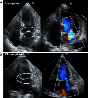
Corte de los tres vasos. Doppler pulsado a nivel del conducto ...
(a) Retrograde left ventricular angiogram demonstrating ventricular ...
Ecocardiograma transtorácico preoperatorio. Jet de insuficiencia ...
Cardiac catheterization showing retrograde filling of the dye to PA ...
Post re-do surgery transthoracic echocardiography -short axis view ...
Transthoracic echocardiography image showing turbulent flow signal from ...
Neoplasms and the Heart | Thoracic Key
Echocardiography in postoperative TOF. a Parasternal short axis view of ...
Echocardiogram. Long axis view with aortic valve (large arrow) and ...
HACEK organisms isolated from definite and probable cases of HACEK ...
Two-dimensional color Doppler echocardiography, showing a turbulent ...
Typical mitral regurgitation associated with SAM and subaortic LVOT ...
(PDF) Feline Hypertrophic Cardiomyopathy: A Spontaneous Large Animal ...
(A) Left ventricular outflow tract obstruction disappeared (Gradients ...
Impact of transcatheter aortic valve replacement on hemodynamic status ...
Echocardiography of Cor Triatriatum showed a membrane structure (arrow ...
Maude PAGÉ | Cardiologist | Hôpital du Sacré-Coeur de Montréal ...
Role of echocardiography for takotsubo cardiomyopathy: clinical and ...
Echocardiogram image in the suprasternal notch position. Color compare ...
A 2D echo with color flow Doppler presents an intraventricular septal ...
Truncus-Arteriosus
Patent-Ductus-Arteriosus-Signs
Persistent-Ductus-Arteriosus
Ductus-Arteriosus-Persistens
Ventricular-Septal-Defect
Patent-Ductus-Arteriosus-Surgery
Atrial-Septal-Defect
Patent-Ductus-Arteriosus-Murmur
Ductus-Arteriosus-and-Ductus-Venosus
Patent-Foramen-Ovale
Patent-Ductus-Arteriosus-and-Newborn
Coarctation-of-the-Aorta
Tortuous-Ductus-Arteriosus
Closure-of-Ductus-Arteriosus
Truncus-Arteriosus-Ultrasound
Ductus-Arteriosus-Location