Please enter url.
Login
Logout
Please enter url.
Ankylosing Spondylitis Mri Si Joints
mavink.com
source
Comments
EPOS™
Osteochondromas of the spine - Clinical Radiology
(PDF) Features of sacral alar fatigue fractures in adolescent athletes ...
Inflammatory bowel disease imaging: Current practice and future directions
Figure 2 from Acetabular paralabral cyst causing compression of the ...
Radiological Imaging Findings of a Case with Vertebral Osteoid Osteoma ...
An uncommon aetiology of cervical radiculopathy: vertebral artery loop ...
Coronal T1 fat suppressed image with percutaneous gadolinium ...
Figure 1 from Hematospermia Evaluation at MR Imaging. | Semantic Scholar
EPOS™
Pustulotic arthro-osteitis (Sonozaki Syndrome): A rare case report ...
The Middle Ear and Mastoid | Radiology Key
MRI evaluation of anal and perianal diseases. - Abstract - Europe PMC
Open management of atraumatic disorders of the sternoclavicular joint ...
Volume 13 Issue 1
Surgical Neurology International
Aguilera-Castro, Ferre-Aracil, Garcia-Garcia-de-Paredes, Rodriguez-de ...
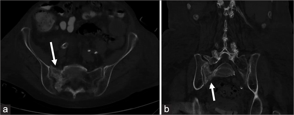
Percutaneous Sacroplasty for Sacral Insufficiency Fractures: Case ...
Magnetic resonance imaging of the pelvis. Urethral tumor (white arrow ...
MR Imaging Evaluation of Pediatric Genital Disorders: - Magnetic ...
Joints: Widened Joint Space | Radiology Key
Recurrent lumbar disc herniation [Neurosurgerypaedia]
Examples of the 5-point grading scale for the PP/EV. A, Grade I: The ...
Lateral Medullary Syndrome and Ipsilateral Hemiplegia (Opalski Syndrome ...
Disc Herniation—Lumbar | Radiology Key
(A) Axial T1-weighted postcontrast fat suppressed spoiled gradient echo ...
Figure 2 from Use of MRI to identify enlarged inferior gluteal and ...
Transperineal injection of hyaluronic acid in the anterior perirectal ...
6 month-old boy a) Contrast-enhanced fat-suppressed spin-echo T1 ...
Pelvic Floor Dysfunction | Radiology Key
Interobserver Agreement of Magnetic Resonance Imaging Signs of ...
Magnetic Resonance Imaging of Hidradenitis Suppurativa: A Focus on the ...
Typical SFA SPACE results: the PAD patient is 72-year-old male with ABI ...
A. An axial T1 weighted MRI of the pelvis showing the right-sided cyst ...
Myelitis | Radiology Key


![Recurrent lumbar disc herniation [Neurosurgerypaedia]](https://neurosurgerypaedia.org/wiki/lib/exe/fetch.php?media=recurrentdischerniation.jpg)