Please enter url.
Login
Logout
Please enter url.
61-year-old female with ruptured distal anterior inferior cerebellar ...
researchgate.net
source
Comments
61-year-old female with ruptured distal anterior inferior cerebellar ...
Image | Radiopaedia.org
(a) NCCT demonstrating HICAS (arrow). (b) CT Carotid Angiogram ...
Patient 5 -Brain CT scan showing infarct in the left temporal lobe ...
Imaging of initial ischemic stroke. (a) CT without contrast, showing ...
Computed Tomographic Determinants of Neurologic Deterioration in ...
On esophagogastroduodenostopy, (A) grade III varices with red wale sign ...
(PDF) Unusual Subdural Hematoma After SPA Use
Brain computed tomography shows epidural hematoma on left temporal area ...
Contrast-enhanced MDCT of neck and thorax, showing partial occlusion of ...
(PDF) Ventricular Pneumocephalus with Meningitis after Lumbar Nerve ...
EPOS™
Non-aneurysmal perimesencephalic subarachnoid haemorrhage | Eurorad
Figure 1 from Macrocephaly and bitemporal arachnoid cysts not ...
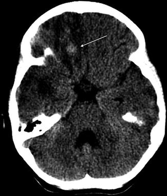
Non-contrast-enhanced axial CT image showing a hyperdense left anterior ...
Subarachnoid Hemorrhage | deemagclinic
Cerebral venous thrombosis in a young female patient with ...
Resolution of "hyperdense" middle cerebral artery post tPA thrombolysis ...
-CT of the head revealed a hyper-dense sign in the right middle ...
Recognizing Some Common Causes of Intracranial Pathology | Radiology Key
CT scan and MRI at admission documented acute obstructive hydrocephalus ...
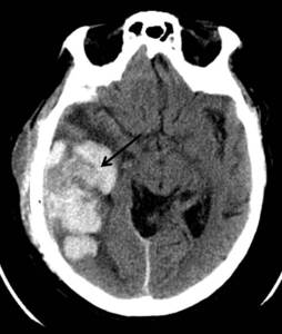
Traumatic Brain Injury | Radiology Key
Interventional Neuroradiology Vasospasm - Los Angeles, CA | Cedars-Sinai
Axial non-contrast CT brain images showing acute haemorrhage into the ...
Radiology MRI: MCA Territory
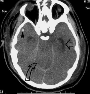
Computed tomography brain showing bilateral cerebellar hypodensities ...
A 60-year-old male presented with sudden-onset severe headache and ...
Cerebral Venous Thrombosis and Multidetector CT Angiography: Tips and ...
CT scan of the head without contrast showing diffuse encephalomalacia ...
A, CT scan of the brain showing diffuse subarachnoid hemorrhage B. CTA ...
Major Pneumocephalus After Lung Resection : A&A Practice
Meningococcal Meningitis: Practice Essentials, Background, Etiology
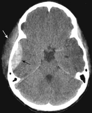
Head and Spine Trauma | Musculoskeletal Key
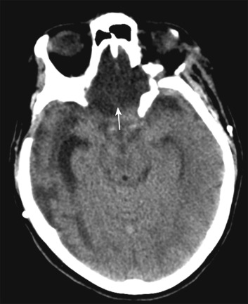
Cranial CT image obtained on the second hospital day at our ...
Axial non contrasted CT of the brain showing subarachnoid hemorrhage in ...