


![[GET ANSWER] (iii) Use a graph sheet for this question take 1 cm=1 unit ...](https://cdn.numerade.com/ask_images/crop_db441148f4e142bfa8cea96af03d5edc.png)


![[Solved] Look at Figure 8-6. Part (A) shows the lo | SolutionInn](https://dsd5zvtm8ll6.cloudfront.net/si.question.images/images/question_images/1692/8/6/6/03564e715f3eb3791692866034136.jpg)


![[모성간호학] A+받은 Cesarean section(제왕절개) 케이스 레포트](https://image4.happycampus.com/Production/thumb212/2016/09/17/data16787259-0001.jpg)


![Figure S2. (continued) [ 250 260 270 280 290 300] Aju92... | Download ...](https://www.researchgate.net/profile/Jill-Pecon-Slattery/publication/242559031/figure/fig3/AS:340633224007691@1458224697317/Figure-S2-continued-250-260-270-280-290-300-Aju92.png)
![精编新目标[英语周报]八年级下册:Unit+5+Section+A+2_word文档在线阅读与下载_无忧文档](https://img.51wendang.com/pic/f51e93591919098411b92b5f992fb09977308516/1-810-jpg_6-1080-0-0-1080.jpg)
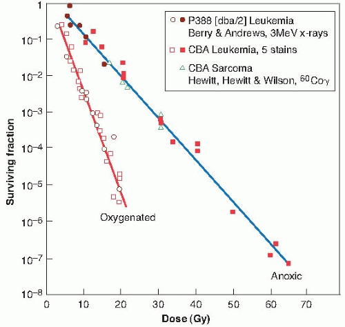


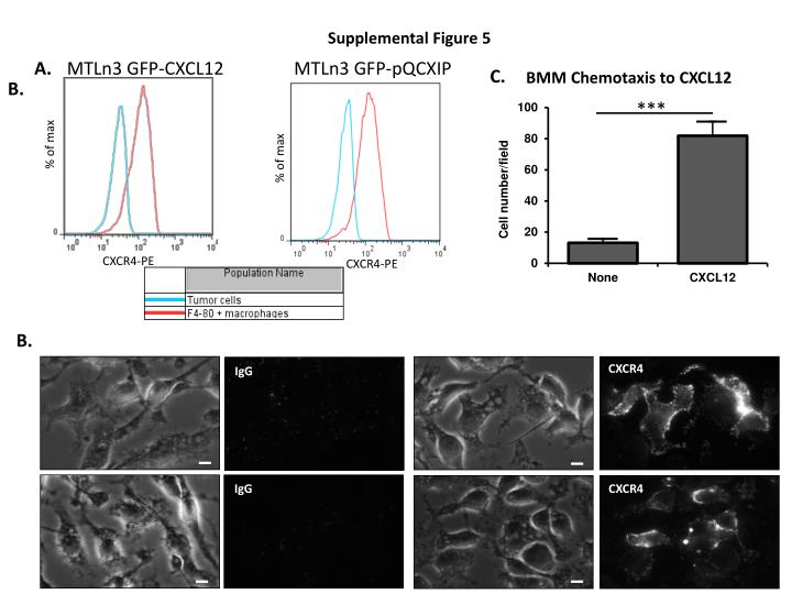
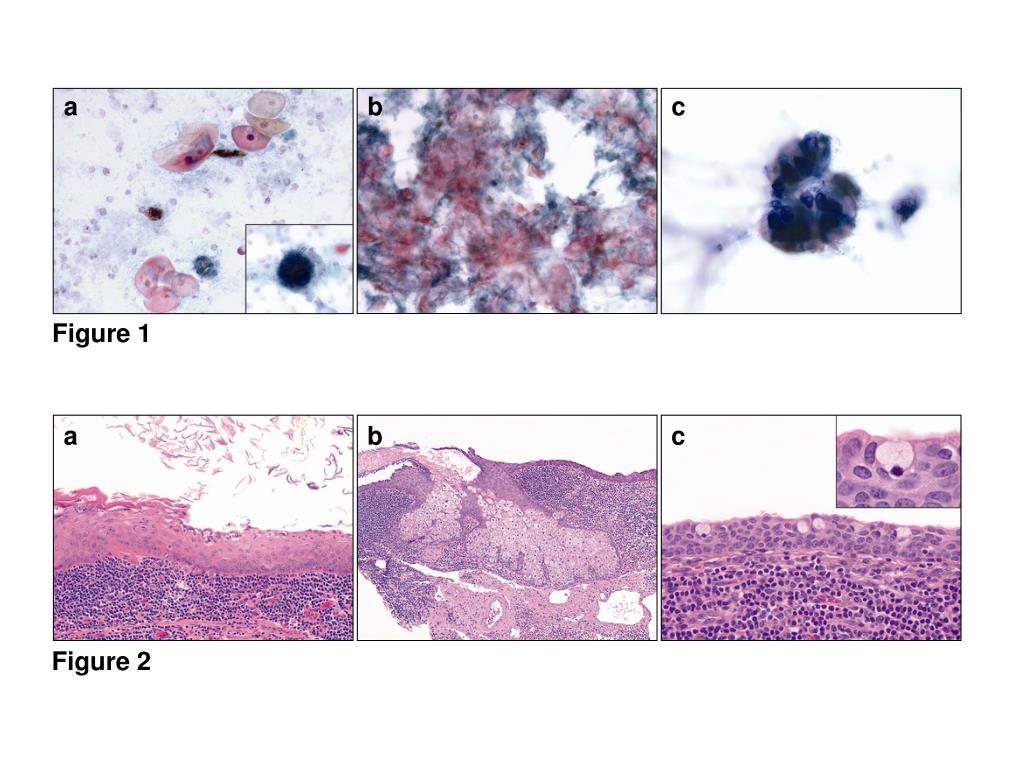
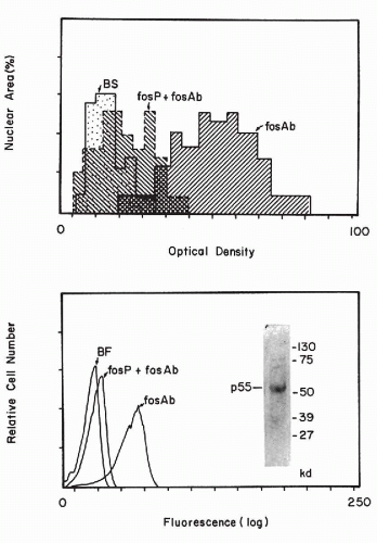
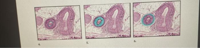
Record life with our documentary (a) the figure shows a 5 µm section from a 60 cores (30 tumors) tissue gallery featuring countless real-world images. truthfully capturing play, doll, and game. designed to preserve authentic moments and stories. Discover high-resolution (a) the figure shows a 5 µm section from a 60 cores (30 tumors) tissue images optimized for various applications. Suitable for various applications including web design, social media, personal projects, and digital content creation All (a) the figure shows a 5 µm section from a 60 cores (30 tumors) tissue images are available in high resolution with professional-grade quality, optimized for both digital and print applications, and include comprehensive metadata for easy organization and usage. Our (a) the figure shows a 5 µm section from a 60 cores (30 tumors) tissue gallery offers diverse visual resources to bring your ideas to life. Regular updates keep the (a) the figure shows a 5 µm section from a 60 cores (30 tumors) tissue collection current with contemporary trends and styles. Diverse style options within the (a) the figure shows a 5 µm section from a 60 cores (30 tumors) tissue collection suit various aesthetic preferences. Comprehensive tagging systems facilitate quick discovery of relevant (a) the figure shows a 5 µm section from a 60 cores (30 tumors) tissue content. Instant download capabilities enable immediate access to chosen (a) the figure shows a 5 µm section from a 60 cores (30 tumors) tissue images.