Please enter url.
Login
Logout
Please enter url.
Kidney Ultrasound
mavink.com
source
Comments
Point of care renal ultrasonography for the busy nephrologist: A ...
Imaging of portal cavernoma cholangiopathy. | Semantic Scholar
Bilateral ureteric calculi – Radiology Cases
Point of care renal ultrasonography for the busy nephrologist: A ...
Contrast-enhanced ultrasound in portal venous system aneurysms: A multi ...
Extra-adrenal Pheochromocytoma Associated With Segmental Renal Artery ...
"Let's Talk About Peritoneal Dialysis" Post 9: Does My Patient Need ...
Emergency imaging in paediatric oncology: a pictorial review | Insights ...
3-year-old male with fulminant hepatic failure, treated with APOLT ...
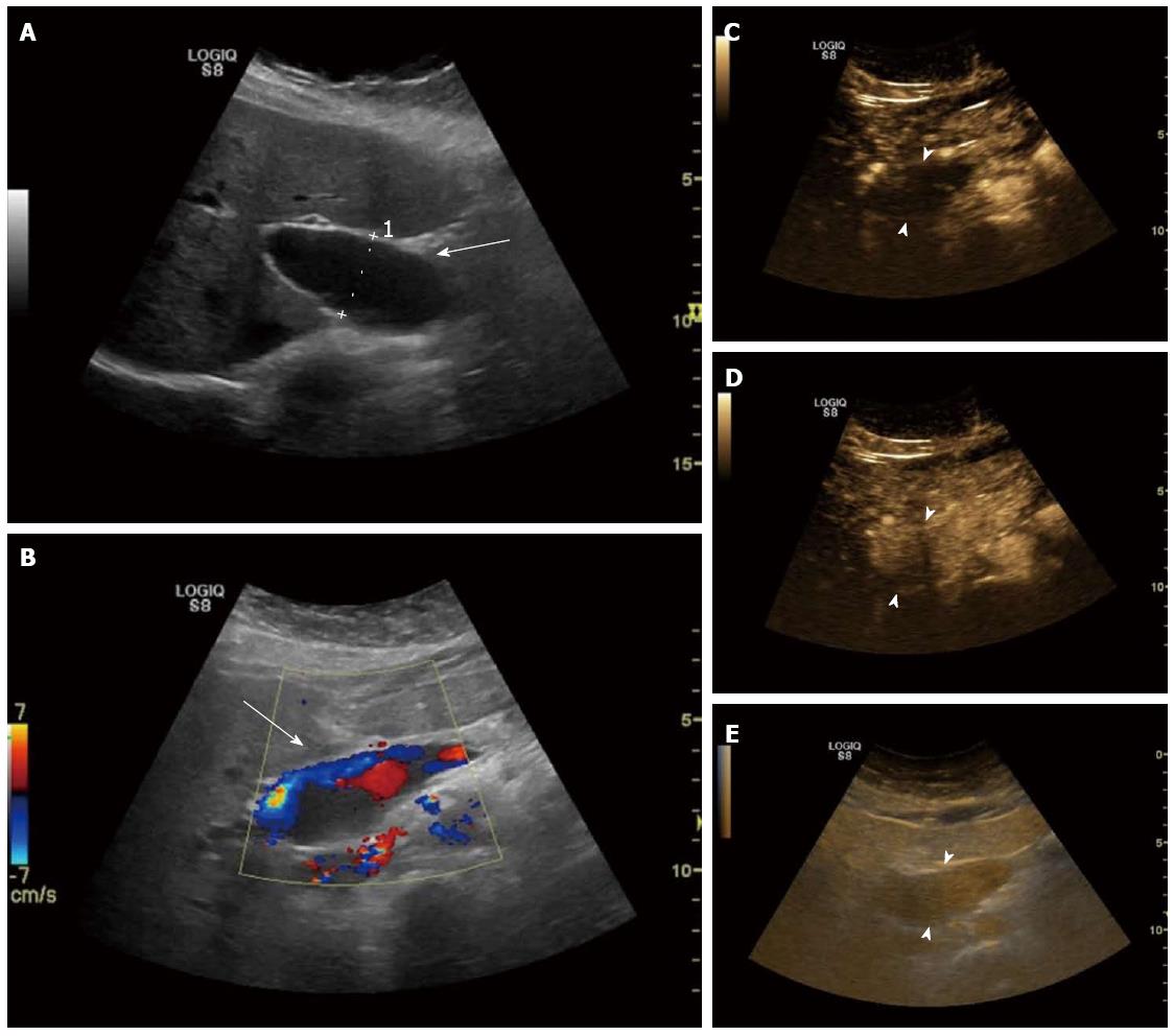
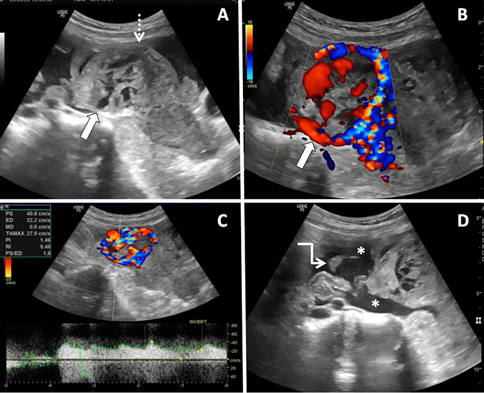
Abdominal ultrasonographic imaging of Case 1. A: Anechoic lesion ...
Medicina | Free Full-Text | Diagnostic Performance of Contrast-Enhanced ...
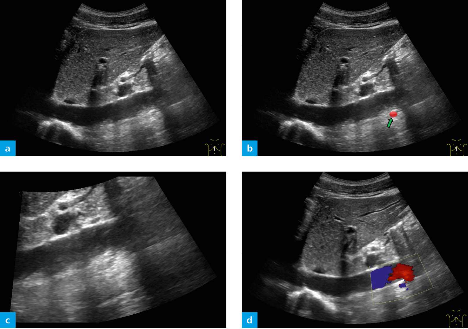
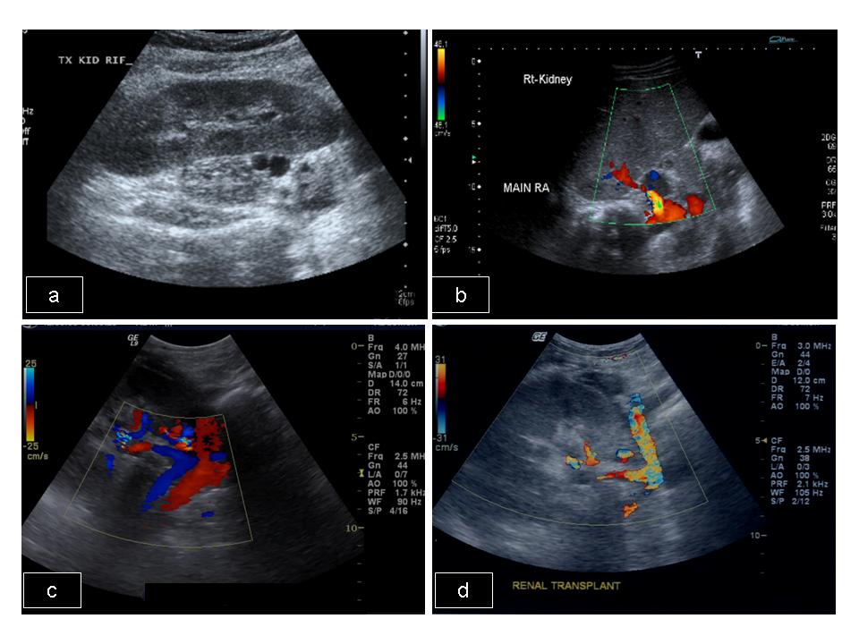
In sagittal B-mode (a) and color Doppler (b), axial B-mode (c) and ...
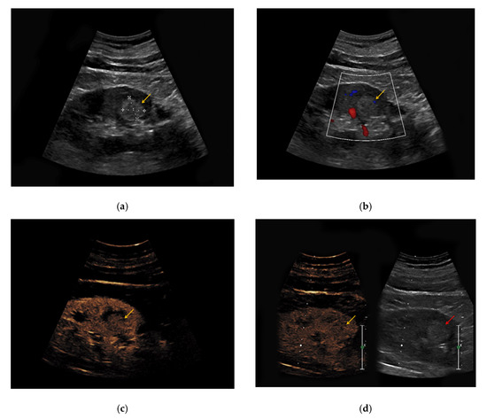
Ultrasound images. (A) The lesion in case 1 was regularly shaped and ...
Color Doppler ultrasonography. Segmental occlusion was shown in the ...
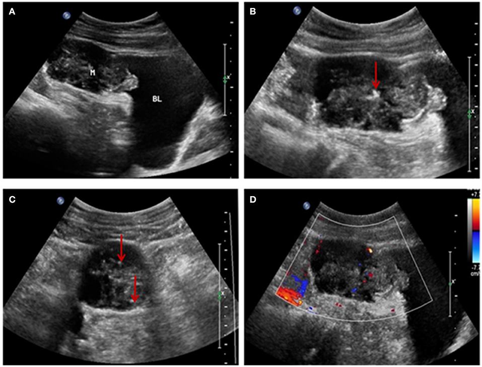
Frontiers | Imaging Features of Urachal Cancer: A Case Report
(PDF) Case Report: Splanchnic Vein Thrombosis as a Complication of ...
arteries | Radiology Key
Cureus | Choriocarcinoma-Driven Uterine Perforation in a Bicornuate ...
EPOS™
Ultrasound Imaging before operation. (a) An uneven slightly higher echo ...
Figure 4 from Sonographic Evaluation of the Abdominal Aorta | Semantic ...
-Ultrasonographic images. (A-C) Ultrasound images show a mixed-density ...
(A) Heterogenous mass at the junction of the pancreatic head & caudate ...
Ultrasound images. (A) The lesion in case 1 was regularly shaped and ...
[PDF] Sonographic Evaluation of the Abdominal Aorta | Semantic Scholar
Persistent fetal lobulation of kidney mimicking renal tumour | BMJ Case ...
Figure 1 from A Rare Case of Accessory Spleen Torsion Diagnosed by ...
Postoperative 3D-CT. An AFX endograft was placed into the Zenith main ...
Angio PLanewave UltraSensitive Imaging (Angio PL.U.S.) as an Innovative ...
Unusual Confluence of Two Rare Syndromes Results in Peculiar Symptoms ...
Turbulent flow in the aortic arch suggests a high cardiac output state ...
Diagnosis and Management of Renal Cystic Disease of the Newborn: Core ...
Nonfetal Imaging During Pregnancy | Radiology Key
Prenatal ultrasound images of the fetus with RTD at 28 +6 wGA showing ...
Figure 1 from Prenatal steroids for microcystic congenital cystic ...
Aorta-Ultrasound
Vascular-Ultrasound
Vascular-Doppler
Ultrasound-of-Abdomen
Bowel-Gas-Ultrasound
Liver-Ultrasound-Scan
Abnormal-Stomach-Ultrasound
Kidney-Scan-Ultrasound
Ultrasound-Abdomen-Complete
Transverse-Liver-Ultrasound
Normal-Stomach-Ultrasound
Ultrasound-Examination
Left-Kidney-Ultrasound
Ultrasound-for-Men
Cancer-Tumor-Ultrasound
Diastasis-Recti-Ultrasound-Images


![[PDF] Sonographic Evaluation of the Abdominal Aorta | Semantic Scholar](https://d3i71xaburhd42.cloudfront.net/6f55974fd0e4dd172360aed18bdf7d71a7bde923/2-Figure1-1.png)