Please enter url.
Login
Logout
Please enter url.
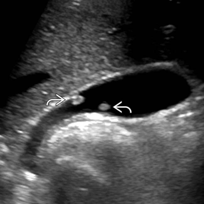
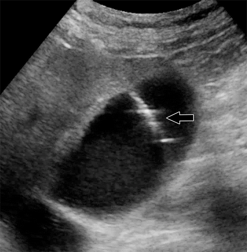
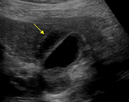
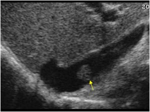
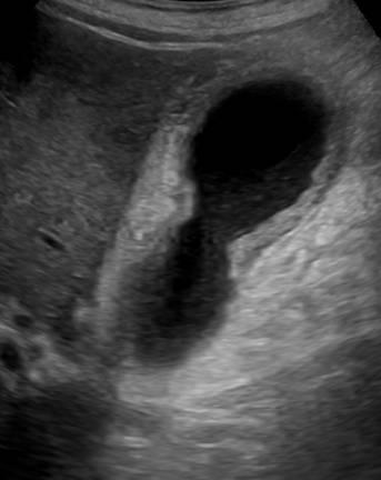
Gallbladder Polyps Ultrasound
mavink.com
source
Comments
Gallbladder Polyps | Radiology Key
Cholesterolosis Vs Adenomyomatosis Ultrasound - A Pictures Of Hole 2018
Intralesional blood vessels in gallbladder lesions on CEUS (arrows). A ...
(PDF) Ultrasound of the gallbladder - is it necessary to fast
Clinical Significance of US Artifacts | RadioGraphics
Adrenal Cysts: Natural History by Long-Term Imaging Follow-Up | AJR
Abdominal Imaging Findings in COVID-19: Preliminary Observations ...
Gallbladder Stones: Imaging and Intervention | RadioGraphics
Lens dislocation | Samantha Salesny MD & Maninder Singh MD | Bronx, NY ...
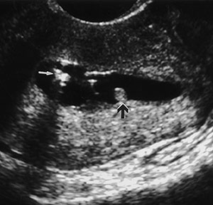
Ultrasonography showing double-linear echogenic structure suggestive of ...
General features of pancreatic cystic neoplasm | Download Scientific ...
Gallbladder and Biliary Tree Flashcards | Quizlet
Abdomen US, day 0 of life (a)–(c). (a) and (b) sagittal images ...
Conventional ultrasound and CEUS images of gallbladder adenoma ...
(a) Hot-Spaxus stent (TaeWoong Medical); (b) Axios stent (Boston ...
Transabdominal ultrasound shows mild focal gallbladder wall thickening ...
Annotated version of Figure 2. The pyloric canal is open. The antrum ...
Abdomen and retroperitoneum | 1.2 Gallbladder and bile ducts : Case 1.2 ...
Abdomen and retroperitoneum | 1.10 Adrenal glands : Case 1.10.6 Adrenal ...
Abdomen and retroperitoneum | 1.2 Gallbladder and bile ducts : Case 1.2 ...
Extra-abdominals arteries or vein: inferior epigastric vessels ...
5.2 Syndromes Flashcards | Quizlet
High-Resolution Sonography for Distinguishing Neoplastic Gallbladder ...
Ultrasound scan of a papillary thyroid non-microcarcinoma (non-PTMC) in ...
abd pathology gallbladder Flashcards | Quizlet
Endometrial Imaging - Part II - myCME
Axial contrast-enhanced CT (left image) of a 62-year-old woman with ...
Three patients with acute interstitial pancreatitis. a 64-year-old ...
Abdomen and retroperitoneum | 1.2 Gallbladder and bile ducts : Case 1.2 ...
Classification of the duplication of the gallbladder. | Download ...
RadioGraphics Update: Contrast-enhanced US Approach to the Diagnosis of ...
Verification of correct central venous catheter placement in the ...
Fetal MRI
Ultrasound findings: gallbladder distension and 1.17 cm wall thickening ...
Coronal (A) and sagittal (B) pelvic male MRI images showing a ...
Gallbladder-Polyps-Ultrasound
Gallbladder-Polyps-Pain
Multiple-Gallbladder-Polyps
Hyperplastic-Gastric-Polyp
What-Is-Gallbladder-Polyp
Gallbladder-Polyps-Symptoms
Cholesterol-Polyps-Gallbladder
Gallbladder-Wall-Polyp
Gallbladder-Polyp-Histology
Hyperplastic-Polyp-Stomach
GB-Polyp-Ultrasound
Symptomatic-Gallbladder-Polyp
Mucosal-Polyp-in-Gallbladder
Gallbladder-Polyp-in-Lumen
Cholesterolosis-Ultrasound
Pictures-of-Stomach-Polyps