Please enter url.
Login
Logout
Please enter url.
Joints | Radiology Key
radiologykey.com
source
Comments
Joints | Radiology Key
Acute wrist pain | Emergency Medicine Journal
The Terry-Thomas Sign - wikiRadiography
Is the Screw in the Joint? - wikiRadiography
(PDF) Ulnar-sided wrist pain. II. Clinical imaging and treatment
Scapholunate ligament Injuries – Fife Virtual Hand Clinic
Abb. 2 8 Clenched-Pencil-Aufnahme: orthograde Abbildung des Spalts ...
(PDF) Differentiating stable buckle fractures from other distal radius ...
Cases | Radiology Key
Functional outcome evaluation using the Gartland-Werley score ...
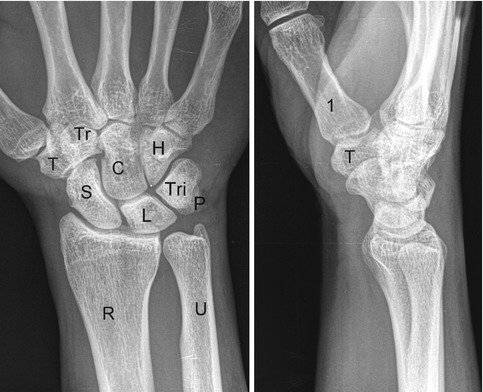
The carpal bones on a lateral plain radiograph of the wrist | The BMJ
The concentric circles method of measuring the ulnar variance. A circle ...
Trauma X-ray - Upper limb - Wrist
Radiographic prediction of lunate morphology in Asians using plain ...
(PDF) Bizarre parosteal osteochondromatous proliferation (Nora's lesion ...
Lunate Dislocation
Middorsal Wrist Pain in the High-Level Athlete: Causes, Treatment, and ...
Fig2:Traumatic recurrent distal radioulnar joint dislocation: a case ...
Negative Ulnar Variance and Kienböck Disease - Journal of Hand Surgery
Reconstruction of Both Volar and Dorsal Limbs of the Scapholunate ...
Radiographic Essentials 2: PA Oblique Wrist Diagram | Quizlet
LearningRadiology
Functional Views of the Wrist - wikiRadiography
49 Scaphoid Nonunion: Medial Femoral Condyle Vascularized Bone Graft ...
Preoperative and Postoperative Imaging of Scapholunate Ligament Primary ...
Image | Radiopaedia.org
Ulnar impaction syndrome after a malunited distal radius fracture with ...
200 A&P ideas in 2022 | anatomy and physiology, nursing study, physiology
Capitate Shortening Osteotomy With Vascularized Bone Grafting for the ...
PA Wrist - Ulnar Deviation Diagram | Quizlet
One year postoperative plain radiograph showed appropriate lunate ...
Adult Distal Radius Fracture Management : JAAOS - Journal of the ...
Wrist | Musculoskeletal Key
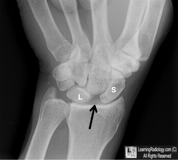
Here the lunate (L) is pie-shaped and dislocated. The schaphoid (S) is ...
Carpal-Tunnel-X-ray-Positioning
Wrist-X-ray-Positioning
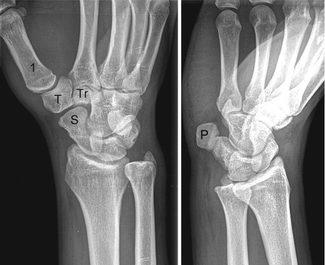
Lateral-Wrist-X-Ray-Pisiform
Navicular-X-ray-Positioning
Scaphoid-View-X-ray-Positioning
PA-Wrist-X-Ray
Lateral-Knee-X-ray-Positioning
Oblique-Elbow-X-ray
Hand-X-ray-Positioning
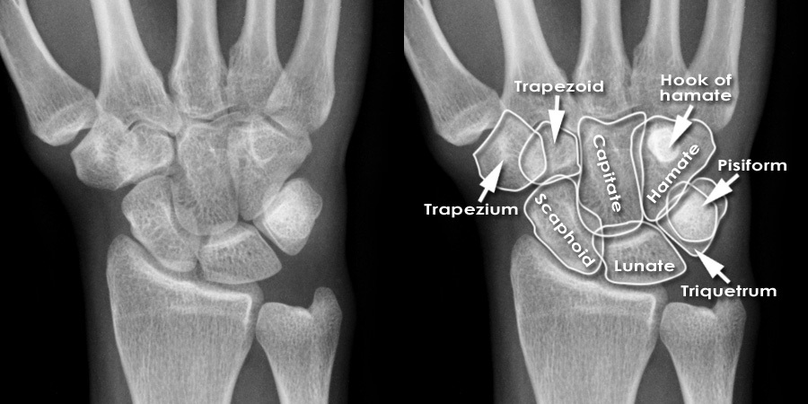
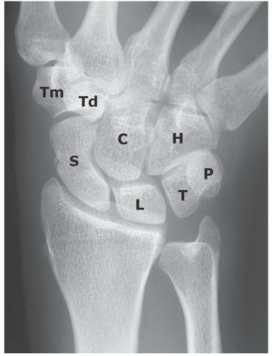
Carpal-Bone-X-ray-Anatomy
Radial-Head-X-ray-Positioning
Standing-Knee-X-ray-Positioning
Pisiform-Dislocation-X-ray
Normal-Wrist-X-ray-Anatomy
Scaphoid-X-ray-Position
AP-Thumb-X-ray-Positioning