Please enter url.
Login
Logout
Please enter url.
Carbon-11-methionine PET ¡mages of 2 patients with advanced ...
researchgate.net
source
Comments
(PDF) Case Report - Accumulation of Tc99m-DMSA-3 in the spleen in a ...
Parathyroid Imaging with Tc-99m Sestamibi Planar and SPECT Scintigraphy ...
Carbon-11-methionine PET ¡mages of 2 patients with advanced ...
FIGURE4. Scintigraphic images ofgranulocytopenic ratswith invasive ...
A 48-yr-old woman with a Warthin's tumor in the right parotid gland ...
In vivo Radionuclide Tests and Imaging - Werner & Ingbar's The Thyroid ...
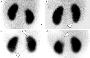
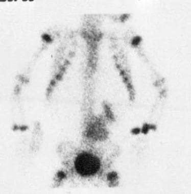
FIGURE1 (Patient9) Anterior and posterior [1231]MIBG imagesof the head ...
a–c [¹⁸F]-FDG-PET/MR image (a), T2-TIRM image (b) and DWI (c) of the ...
Bone-Forming (Osteogenic) Lesions | Radiology Key
FIGURE2. A 79-yr-oldwoman withpapillarythyrokicarcinoma The poste ...
(Left) Baseline ""Tc-DTPA renography shows more delayed drainage of the ...
Overall confidence levels before and after DaTscan. | Download ...
Technetium-99m-Hexamibi (left panel) and @°@T1 (right panel) SPECT ...
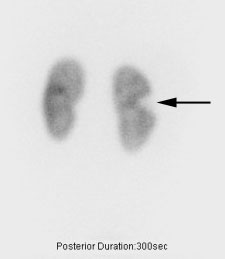
Static renal technetium-99 m dimercaptosuccinic acid (DMSA ...
Molecular Breast Imaging in Breast Cancer Screening and Problem Solving ...
Figure 1 from Iodine-123-MIBG SPECT versus planar imaging in children ...
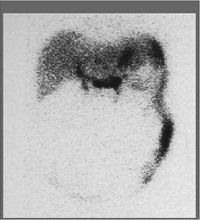
Untitled Document [stritch.luc.edu]
Semiquantitative analysis of power doppler ultrasonography versus Tc ...
FiGURE2. Apophyseal joint disease above a solid lateral LA-Si fusion9 ...
Blood-pool images in plantar (top left), lateral (top right), an terior ...
FIGURE3 Buddâ€"Chiari syndrome:typical scintigraphicpattern. On 10/23/1 ...
Whole-body images of rab bits acquired 20 hr after intravenous in ...
Dual-phase Tc-99m MIBI planar scintigraphy. The early images (a) show ...
Neuroblastoma Imaging: Practice Essentials, Radiography, Computed ...
FIGURE a Anterior image oftheabdomen (A)24and(B)30hrafter injection of ...
Gastrointestinal and Correlative Abdominal Nuclear Medicine Imaging ...
Lymphoscintigraphy (anterior and posterior images) of lower limb (LL ...
Non-Hodgkin's disease (Tcell) ina2-yr-old boy. (A) Anteriorview and (B ...
Anterior (left) and posterior (right) planar views obtained 7 days ...
Gallbladder ejection fraction in Case 2 following compounded sincalide ...
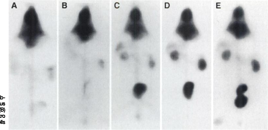
Pet-1 expression in brain is restricted to serotonergic neurons. A , B ...
Gallery: Image (444)
Figure 1 from Blood-pool imaging using technetium-99m-labeled liposomes ...
Case 11. (a) Anterior and (b) posterior scans of the thoracic area ...


![FIGURE1 (Patient9) Anterior and posterior [1231]MIBG imagesof the head ...](https://www.researchgate.net/profile/Roel-Hoogma/publication/20993494/figure/fig2/AS:667801933066259@1536227796599/FIGURE1-Patient9-Anterior-and-posterior-1231MIBG-imagesof-the-head-do-not-revealany_Q320.jpg)
![a–c [¹⁸F]-FDG-PET/MR image (a), T2-TIRM image (b) and DWI (c) of the ...](https://www.researchgate.net/publication/352975841/figure/fig1/AS:1041717633499136@1625376246230/a-c-Upper-neck-level-II-on-T2-TIRM-transversal-a-F-FDG-PET-b-and-DWI-at_Q320.jpg)
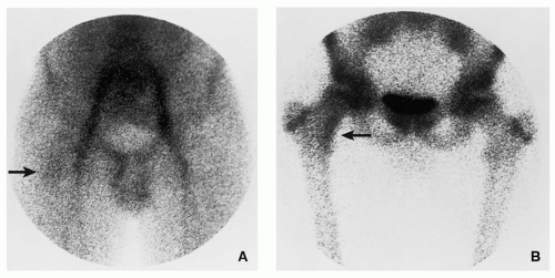


![Untitled Document [stritch.luc.edu]](https://stritch.luc.edu/lumen/meded/radio/curriculum/medicine/GI_bleed.jpg)