Please enter url.
Login
Logout
Please enter url.
Ankylosing Spondylitis Mri Si Joint
mavink.com
source
Comments
Ankylosing Spondylitis - Radsource
C5 pure motor spinal cord injury: A case with a rare manifestation of ...
An algorithmic strategy for selecting a surgical approach in cervical ...
(PDF) Cervical disc herniation causing Brown-Sequard syndrome: Case ...
Four representative cases showing the features of degenerative ...
Figure 3 from A single-stage posterior approach with open reduction and ...
Preoperative anteroposterior and lateral X-ray of a 39-year-old female ...
Magnetic resonance imaging scans (T2-weighted) of the spine in the (a ...
Figure 1 from The clinical effect of percutaneous kyphoplasty for the ...
Ankylosing Spondylitis | Radiology Key
Bilateral Facet Dislocation
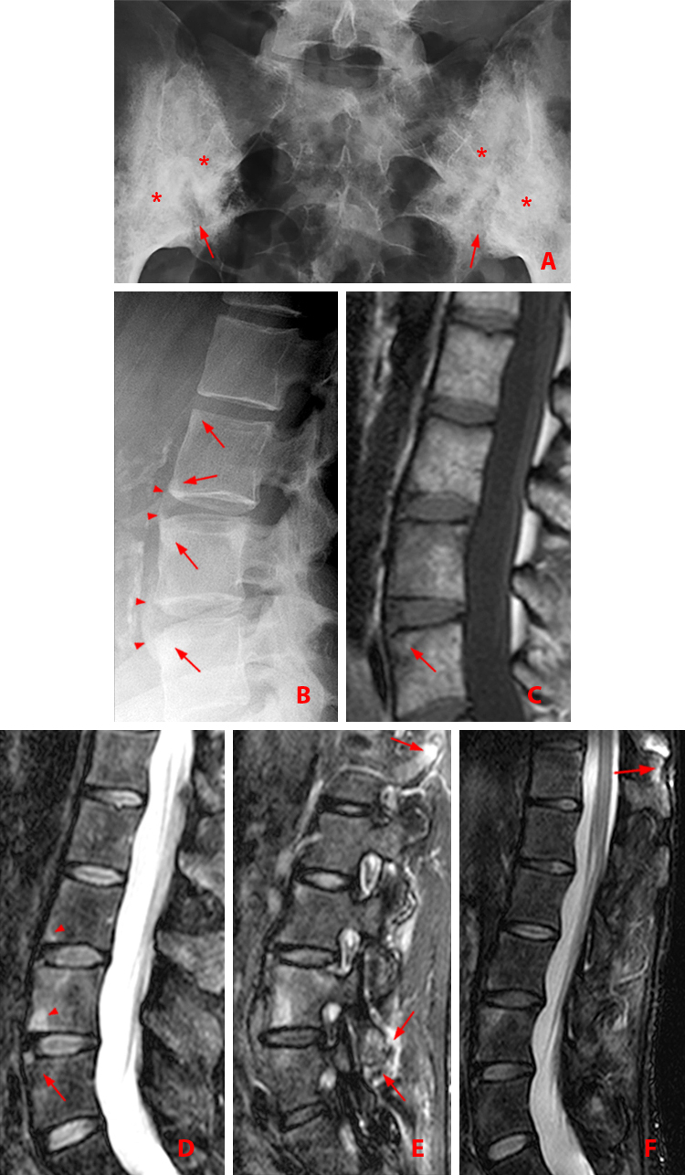
Pediatric Spinal Infections (Chronic) | Neupsy Key
Nurick classification system for degenerative cervical myelopathy ...
Coexisting cervical OPLL and thoracic OLF. | Download Scientific Diagram
Ankylosing Spondylitis - Radsource
Customizable, effective braces for scoliosis and spondylosis in ...
MR Image; Discitis After Nucleoplasty Radiofrequency of L4-L5 ...
Vertebral Lesions: Imaging | Neupsy Key
Optimizing the Imaging Options | Radiology Key
[PDF] Charcot spinal arthropathy: an increasing long-term sequel after ...
Ankylosing Spondylitis | Radiology Key
Case 2 A 69-year-old male patient presented with numbness in his ...
Cervical PsA. (a) Lateral radiographs in the neutral position and (b ...
Percutaneous Ozone Treatment for Herniated Lumbar Discs: 1-Year Follow ...
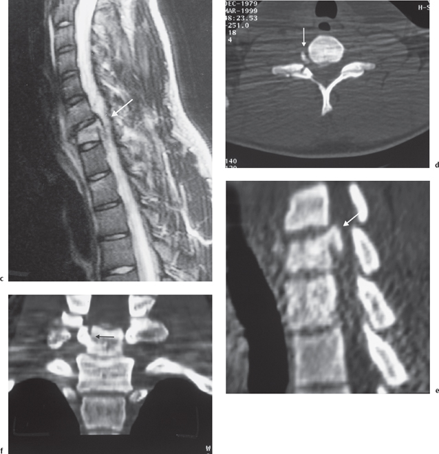
Cervical spine CT (left) and MRI (right). Extensive OPLL extending from ...
Clinical characteristics and surgical outcome of thoracic myelopathy ...
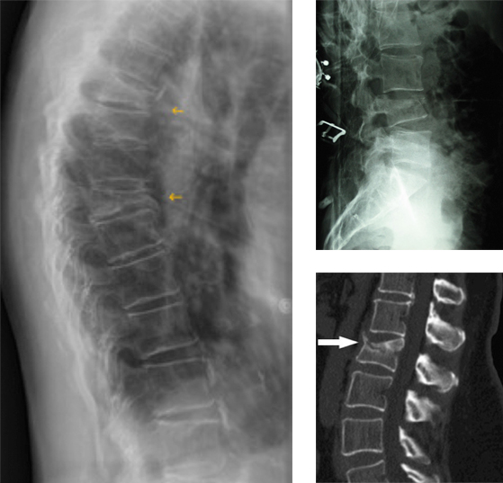
X-ray images of vertebral compression fracture: a) x-ray images of ...
A±C Sagittal and axial MR images of the thoracic spine. A Sagittal ...
Common Spine Disorders Associated with Neck Pain | Radiology Key
(a) The direct X-ray lateral graphy imaging of the cervical spine ...
Maximum intensity projection image of 18F-FDG PET/CT scan (A); PET, CT ...
(A, B) Frontal and lateral X-rays show inferior vena cava sign at L1 ...
a Case 7: male, 52 years old. Preoperative MRI: severe DDD at L5–S1 and ...
Anterior lumbar discectomy and fusion for acute cauda equina syndrome ...
Ankylosing-Spondylitis-Spine-MRI
People-with-Ankylosing-Spondylitis
Ankylosing-Spondylitis-Radiopaedia
Severe-Ankylosing-Spondylitis
Cervical-Ankylosing-Spondylitis
Ankylosing-Spondylitis-Eyes
Ankylosis-Spine
Ankylosing-Spondylitis-Lumbar-MRI
Sacroiliitis-MRI
X-ray-of-Ankylosing-Spondylitis
Advanced-Ankylosing-Spondylitis
Ankylosing-Spondylitis-Photos
Juvenile-Ankylosing-Spondylitis
Stages-of-Ankylosing-Spondylitis
Ankylosing-Spondylitis-MRI-Pelvis
Ankylosing-Spondylitis-Severe-Pain


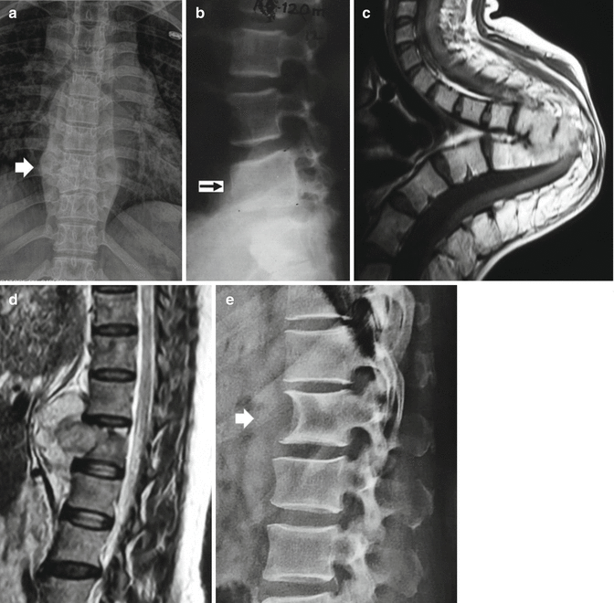


![[PDF] Charcot spinal arthropathy: an increasing long-term sequel after ...](https://d3i71xaburhd42.cloudfront.net/17c7a55e59e0047cba41b73aa93c6ad9f101388b/3-Figure1-1.png)