Please enter url.
Login
Logout
Please enter url.
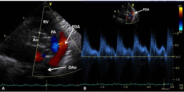
Patent Ductus Arteriosus Echo
ar.inspiredpencil.com
source
Comments
Echocardiographic Evaluation of Neonates with Suspected Heart Disease ...
Repeat mitral valve repair for haemolysis in children | Thoracic Key
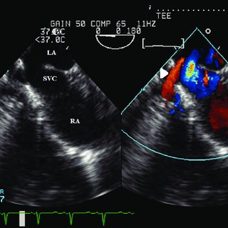
View of the superior sinus venosus defect between the SVC and LA with ...
Ascending aorta aneurysm upper sinotubular junction visualised by TEE ...
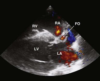
Transthoracic echocardiography. Apical four chamber view demonstrating ...
Transthoracic echocardiography image showing turbulent flow signal from ...
(PDF) Hypoplastic right heart syndrome, absent pulmonary valve, and non ...
A parasternal long-axis view showing a truncus arteriosus (TA), a ...
(PDF) Supraventricular Tachycardia and Tricuspid Regurgitation in the ...
Electrocardiogram on presentation showing sinus tachycardia with a ...
Measurement of the ejection fraction using M-mode in the parasternal ...
Vegetation on tricuspid valve – echocardiographic image – Cardiophile MD
Sinus of Valsalva with thrombus as seen on parasternal short axis view ...
Sonographer’s Perspective of Evaluating Diastolic Function | Thoracic Key
Intramural hematoma due to a penetrating aortic ulcer (arrow) is shown ...
Multiple Asymptomatic Tricuspid Papillary Fibroelastomas - Case Report
Color Flow Doppler Saturation Ultrasound | Postgraduate educational ...
Cardiac murmurs: congenital heart disease | Veterian Key
post-opératoire (3 mois): coupe parasternale grand axe, mode ...
Left atrial septal pouch thrombus seen by color Doppler imaging during ...
Transthoracic echocardiogram from a modified apical window showing ...
Abnormalities of Left Ventricular Outflow - Echocardiography in ...
(A) Left ventricular outflow tract obstruction disappeared (Gradients ...
Transthoracic echocardiography demonstrates severe aortic regurgitation ...
TTE showing the defect in the upper part of the septum | Download ...
Artifacts, and Quality control Time (2 Hr)Central standard time
Transthoracic echocardiography: Apical 2-chamber view with color ...
Unbalanced common atrioventricular canal after the Fontan operation-a ...
Echocardiogram in the short-axis view of aortic valve level showing ...
Site of transseptal puncture. LAA: Left atrial appendage, PVI ...
Echocardiographic images of the large muscular ventricular septal ...
(a) Apical five-chamber view with colour Doppler showing severe ...
Pathological specimens showing various patterns of ventricular ...
Holodiastolic flow reversal was seen in the proximal descending ...
Four-chamber apical approach showing mitral valve regurgitation ...