Please enter url.
Login
Logout
Please enter url.
Parotid Gland Ultrasound Measurement
mavink.com
source
Comments
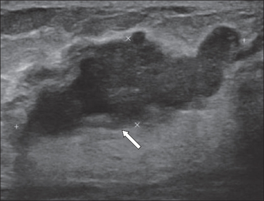
Lymphatic cystadenoma (Warthin tumor). Typical fluid spaces present in ...
35 year old female diagnosed with right sided hydrocele of the canal of ...
Echogenic foci associated with malignancy. 70-year-old female with ...
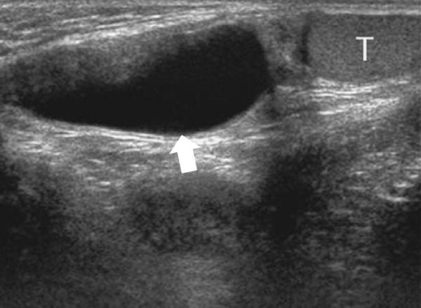
Liquefied hematoma (arrows, anechoic fluid collection). | Download ...
A 54-year-old woman diagnosed with papillary thyroid carcinoma. A 7 mm ...
Synovitis | Musculoskeletal Key
Longitudinal image of a hypoechoic nodule with a spiculated margin ...
Clinics in diagnostic imaging (180) | SMJ
Canal of Nuck Abnormalities in Pediatric Female Patients | RadioGraphics
Hip and Thigh Ultrasound | Clinical Gate
Malignancy Risk Stratification of Thyroid Nodules: Comparison between ...
Photograph of a zigzag incision in the wrist. An intraneural ganglion ...
BIR Publications
A 27-year-old woman with gradual increase in a thyroid colloid cyst ...
Ultrasonographic evaluation of cholecystoduodenostomy sites in six cats ...
Ultrasonographic features of second nodal recurrences (6 years after ...
Photomicrograph showing metastatic parathyroid carcinoma to the lymph ...
35 year old female diagnosed with right sided hydrocele of the canal of ...
Supraclavicular Block Labeling Diagram | Quizlet
Normal ultrasound anatomy and common anatomical variants of the thyroid ...
US image of the right groin shows herniation of the ovary (black ...
a. Pure cyst 5 mL, before PEI. b. Solid residue 0.2 mL, 6 months after ...
Correlation between TIRADS and ATA grading systems and Bethesda ...
Figure 8 from The role of sonography in thyroid cancer. | Semantic Scholar
Lipoma in a 54-year-old woman who complained of a palpable nodule in ...
US image shows the left ovary (arrows) displaced toward the opening of ...
IgG4-related Disease from Head to Toe | RadioGraphics
Round-shaped, hypoechoic, cervical lymph node in a patient affected by ...
The same patient as in Fig. 1. The arrow shows the target lymph node ...
Medullary Thyroid Carcinoma: Imaging | SpringerLink
16 Male Genital Tract | Radiology Key
Ultrasound as a Localization Technique in Hyperparathyroidism ...
Longitudinal ultrasound through the suprapatellar recess of the right ...
Elbow Ultrasound | Radiology Key
Liposarcoma: low-grade myxoid recurrent. Ultrasound image (A) shows ...
Parotid-Gland-On-Ultrasound
Submandibular-Gland-On-Ultrasound
Salivary-Gland-Disease
Sjogren-Salivary-Gland
Salivary-Gland-Stone-Ultrasound
Parotitis-Ultrasound
Parotid-Duct-Ultrasound
Salivary-Gland-Mass
Parotid-Gland-Biopsy
Sublingual-Gland-Ultrasound
Parotid-Abscess-Ultrasound
Normal-Parotid-Ultrasound
Salivary-Glands-in-Neck
Sialadenitis-Ultrasound
Parotid-Ultrasound-Protocol
Salivary-Glands-Anatomy-Image