Please enter url.
Login
Logout
Please enter url.
Figure 2 from A Rare Presentation with Angina and Pseudoinfarct Ecg ...
semanticscholar.org
source
Comments
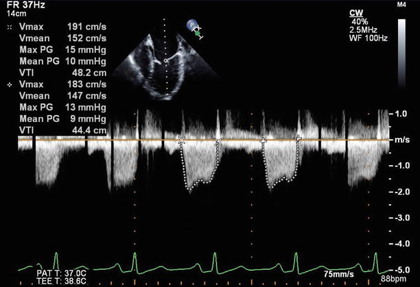
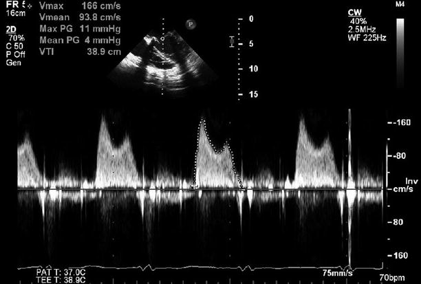
Continuous wave Doppler signal across the tricuspid value showing low ...
Low Transvalvular Pressure Gradients 14 Days After the Implantation ...
Rheumatic Mitral Valve Disease | Radiology Key
Severe Functional Tricuspid Stenosis Secondary to a Giant Saphenous ...
Mitral Valve Area Calculator - CALCULATORVGW
Patent Ductus Arteriosus and Aortopulmonary Window | Thoracic Key
Doppler Recognition of Low or Normal Central Venous Pressure from ...
Practical Echocardiography Cases: Video Index
Two-dimensional midesophageal long axis transesophageal... | Download ...
Aortic Valve Anatomy and Embryology | Thoracic Key
Analyze This Image: Make The Diagnosis in Vein
9 Continuous-wave Doppler imaging confirms mitral regurgitation ...
Sanjay Banypersad and Keith Pearce: Succesful Accreditation in ...
Figure 3 from Use of a melody pulmonary valve in transcatheter valve-in ...
Holodiastolic Flow Reversal In The Distal Portion Of The Aortic Arch ...
JCM | Free Full-Text | Ultrasound Assessment in Cardiogenic Shock ...
Shows TDI of LV lateral wall (systolic and diastolic wave). | Download ...
Diastolic and Systolic Cardiac Dysfunction in Pectus Excavatum ...
Left Atrial Appendage, Intraoperative Echocardiography, and the ...
Continuous-wave Doppler with Valsalva maneuver on TTE in the apical ...
Tricuspid Regurgitation Velocity Unreliable Indicator of Pulmonary ...
Echocardiogram showing diastolic hepatic vein flow reversal. | Download ...
Dr Louise Barnett BVM BVS(Hons) PGCert (SAMS) MANZCVS (SA Radiology)
Unilateral Pulmonary Edema Secondary to Mitral Valve Perforation ...
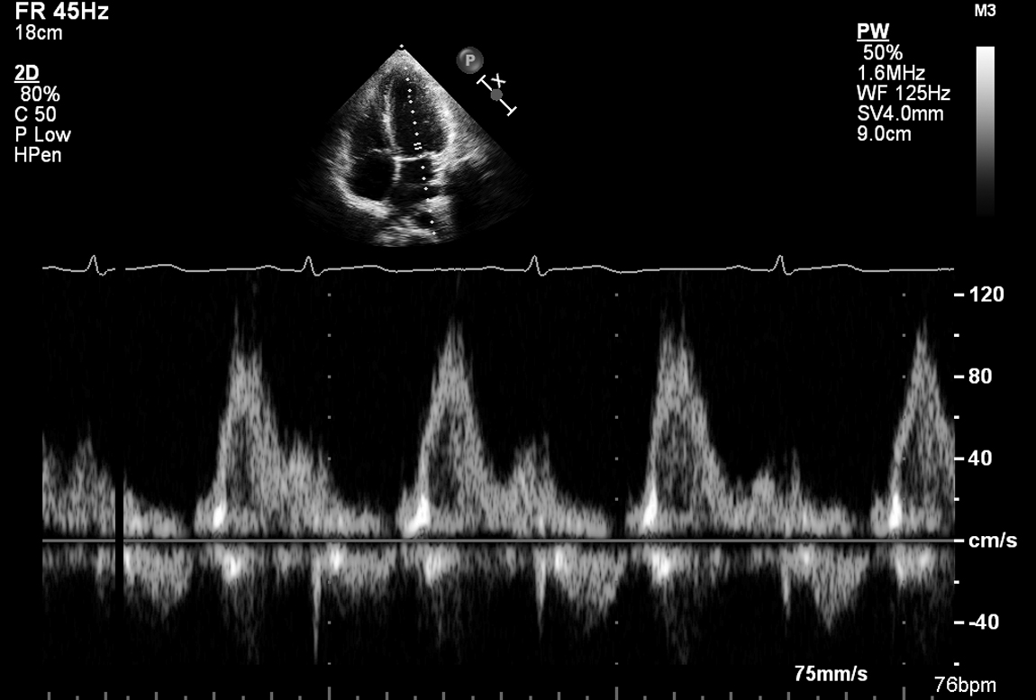
Pulsed-wave Doppler of the left superior pulmonary vein showing ...
Analyze This Image: What Is Being Shown Here? - Page 2
Transthoracic echocardiography apical four-chamber view showing ...
Infective Endocarditis | Radiology Key
TCTAP C-232 Wrong Destination of a Trans-Septal Puncture Needle, What ...
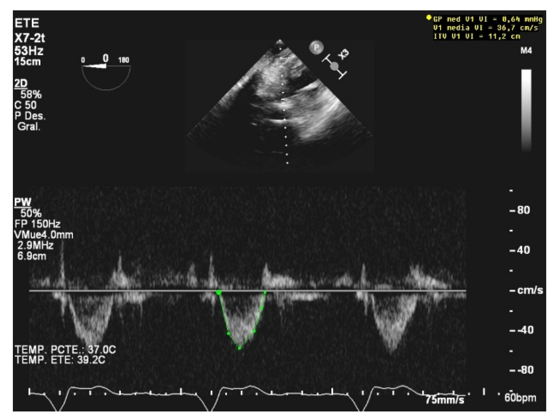
Relación S/D 39,5%/57,2: menor a 1 (0,69). | Download Scientific Diagram
The Atria Are Stunned, but You Shouldn't Be
Ultrasound Machines - "Knobology" — Taming the SRU
Figure 3 from Recurrent Hemopericardium With Cardiac Tamponade as an ...
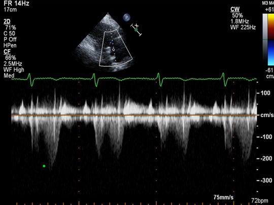
Pulse wave doppler over ASD showing bidirectional shunt. | Download ...
Gradient across Pulmonary Valve before BPV. | Download Scientific Diagram