Please enter url.
Login
Logout
Please enter url.
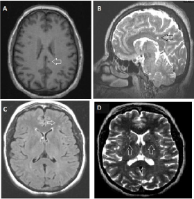
Marchiafava–Bignami disease-like lesions due to central nervous system ...
casereports.bmj.com
source
Comments
Combined susceptibility effect and T1-weighted hyperintensity were seen ...
Axial magnetic resonance imaging in the posterior reversible ...
Differential Diagnosis of Bilateral Thalamic Lesions (PDF Download ...
(PDF) Neuroimaging findings of linear scleroderma of the head and face ...
A 15-year-old boy with a pineal region germinoma, proved by surgical ...
Quantitative Electroencephalographic Changes and Retinal Alterations in ...
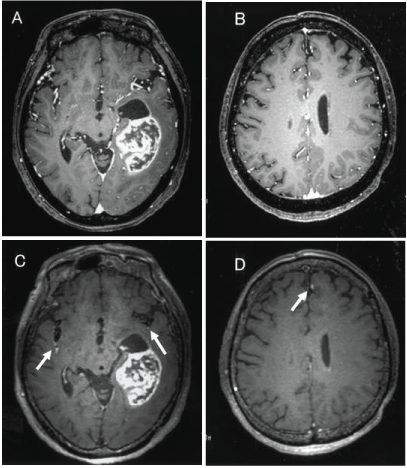
Artifacts secondary to the shunt reservoir. The SS-GRE MR image is the ...
Brain Magnetic Resonance Imaging Abnormalities in Adult Patients With ...
A 3-month-old boy with abusive head trauma. a An axial... | Download ...
Detectability of Intraaxial Lesions and Disseminations for Primar
Monkeypox encephalitis with transverse myelitis in a female patient ...
(PDF) Spinocerebellar Ataxia Type 31 with Blepharospasm
Placental Pathology in Neonatal Stroke: A Retrospective Case-Control ...
T2 weighted MRI scans showing diffuse hyperintense signal abnormalities ...
Magnetic resonance imaging of brain showing high T2 signal in bilateral ...
Cerebral sinovenous thrombosis in pediatric practice | SpringerLink
Postoperative MRI. (a) T2, (b) Flair, (c) T1 without and (d) T1 with ...
Frontiers | Challenges in Drug Discovery for Neurofibromatosis Type 1 ...
Axial magnetic resonance images of the brain showing (A)... | Download ...
Patient F5 (P1) MRI, age 4 years 2 months (A-D), and 7 years 6 months ...
Neuroimaging of CNS infection in haematological malignancy: important ...
Case 1: a Coronal noncontrast CT image from HD 25, showing a 2 cm ...
Figure 1 from Teaching NeuroImages: Acute crossed cerebellar diaschisis ...
The sequences of the relevant areas of the ARFGEF2 gene are depicted ...
Cephaloceles | SpringerLink
A 41-year-old male with pulmonary Koch's with meningitis. Magnetic ...
(a) Brain CT scan showing hypodense lesions in subcortical white matter ...
Typical brain imaging changes after infection of EV-A71. (A) Lesion in ...
Globus pallidus-related MRI findings of PKAN. | Download Scientific Diagram
| Brain MRI findings in MNGIE. MRI of MNGIE patient at age 16 with ...
Systemic vascular phenotypes of Loeys-Dietz syndrome in a child ...
An 8-month-old boy with abusive head trauma. a An axial... | Download ...
Initial Magnetic Resonance Imaging (MRI) Findings Showing Abnormal ...
| Detection of axonal injury with conventional magnetic resonance ...
Central-Nervous-System-Vasculitis
Lupus-Brain
Vasculitis-MRI
CNS-Vasculitis
Lupus-Brain-Lesions
Lupus-Neuropathy
Nervous-System-Diseases-List
Central-Nervous-System-Nerves
SLE-Vasculitis
Systemic-Lupus
Cerebral-Vasculitis
Lupus-Myositis
Central-Nervous-System-Real
Lupus-Cells
Systemic-Lupus-Erythematosus-Disease
Lupus-Cerebritis