Please enter url.
Login
Logout
Please enter url.
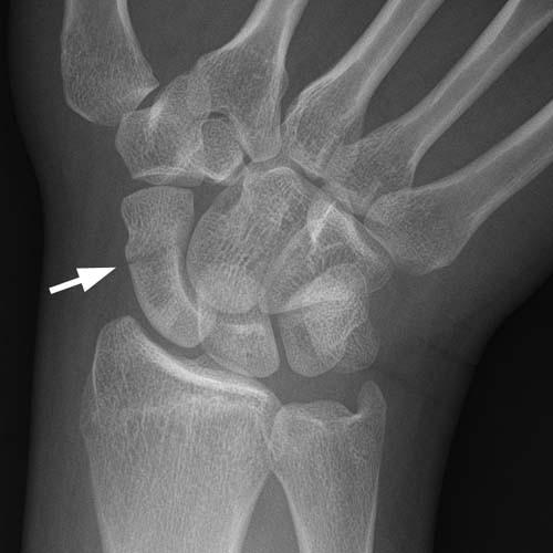
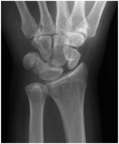
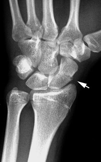
On a normal AP wrist the lunate (L) has a nice rhomboid shape ...
researchgate.net
source
Comments
Transscaphoid perilunate dislocation – ScienceOpen
(a) CT-scan showing consolidated fracture of the scaphoid and ...
Schematic representation of a normal wrist with normal parallel zigzag ...
Wrist | Radiology Key
Scaphotrapeziotrapezoidal Joint Arthritis - WikiSM (Sports Medicine Wiki)
Divergent Fracture–Dislocation of the Hamatometacarpal Joint: Case ...
Avascular necrosis of lunate bone: Kienbock disease - The American ...
Imaging Scaphoid Fractures - wikiRadiography
(PDF) An unusual case of gout in the wrist: The importance of ...
Negative and neutral ulnar variance. A: In negative ulnar variance, the ...
Radiodiagnosis - Imaging is Amazing-Interesting cases: Scapholunate ...
Wrist fracture - MEDizzy
Scaphoid Fracture - Orthopedics - Medbullets Step 2/3
X-Wrist
Soft Tissue Calcification and Ossification | Radiology Key
Hamate fracture - WikEM
Orthopedic | Anesthesia Key
(PDF) Pisiform–Hamate Coalition With Entrapment Neuropathy of the Deep ...
Scaphoid fracture locations. a-c Posteroanterior radiographic wrist ...
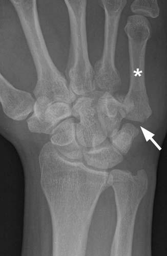
Here the lunate (L) is pie-shaped and dislocated. The schaphoid (S) is ...
Hand | Radiology Key
Wrist and hand | Musculoskeletal Key
Medoff Wrist Fixation System
Wrist Trauma Radiographic Evaluation - Hand - Orthobullets
X-Wrist
Imaging Scaphoid Fractures - wikiRadiography
Radiographic PA view of the wrist shows a soft tissue mass (white ...
Posteroanterior radiograph of the left wrist showing positive ulnar ...
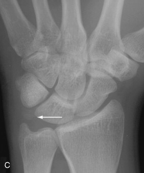
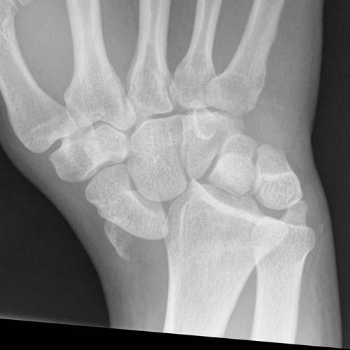
PA radiograph of the injured right wrist demonstrating normal ...
CE4RT - Radiographic Positioning of the Wrist for X-ray Technologists
Wrist and Hand | Radiology Key
Functional Views of the Wrist - wikiRadiography
Forearm, Elbow, and Upper Arm | Radiology Key
Frontal radiograph demonstrating lunate malacia. The lunate is ...
Osteochondroses - wikiRadiography