Please enter url.
Login
Logout
Please enter url.
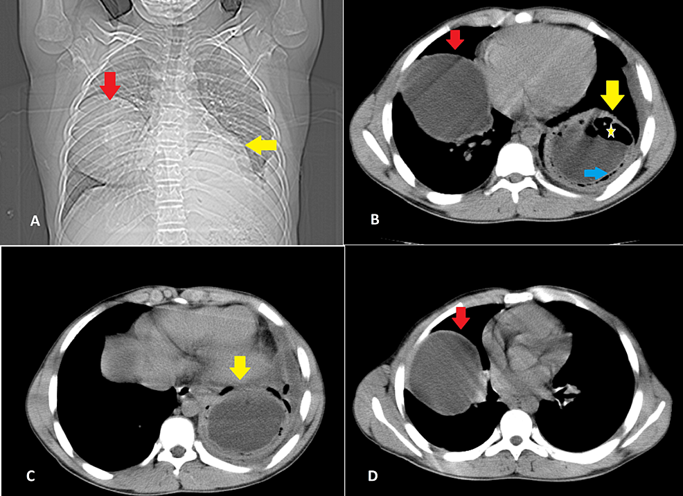
Cureus | Seronegative Bilateral Pulmonary Hydatid Cysts in a 15-Year ...
cureus.com
source
Comments
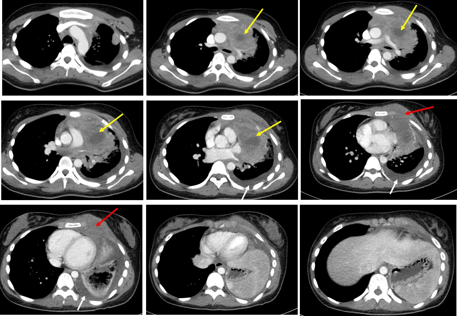
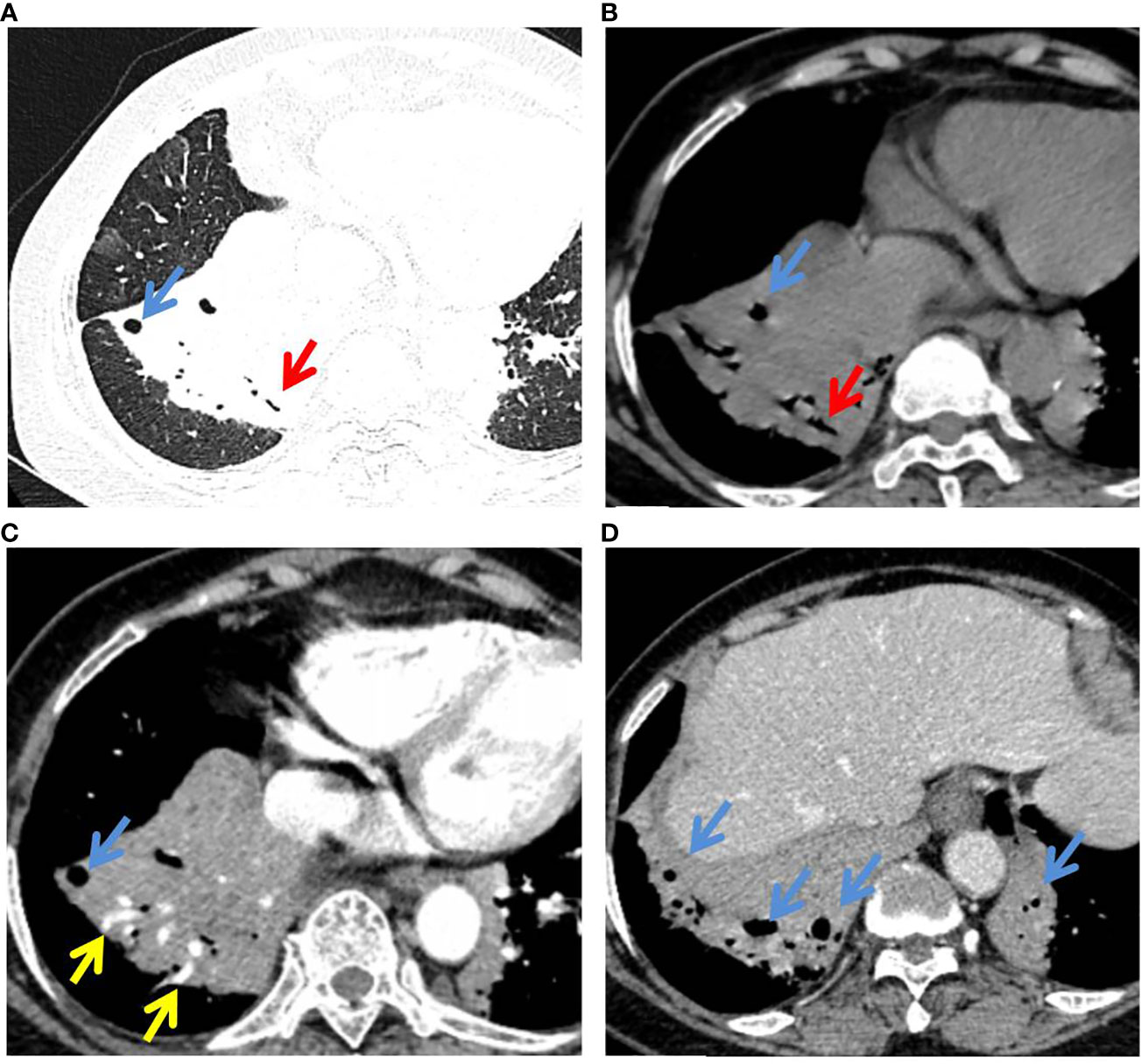
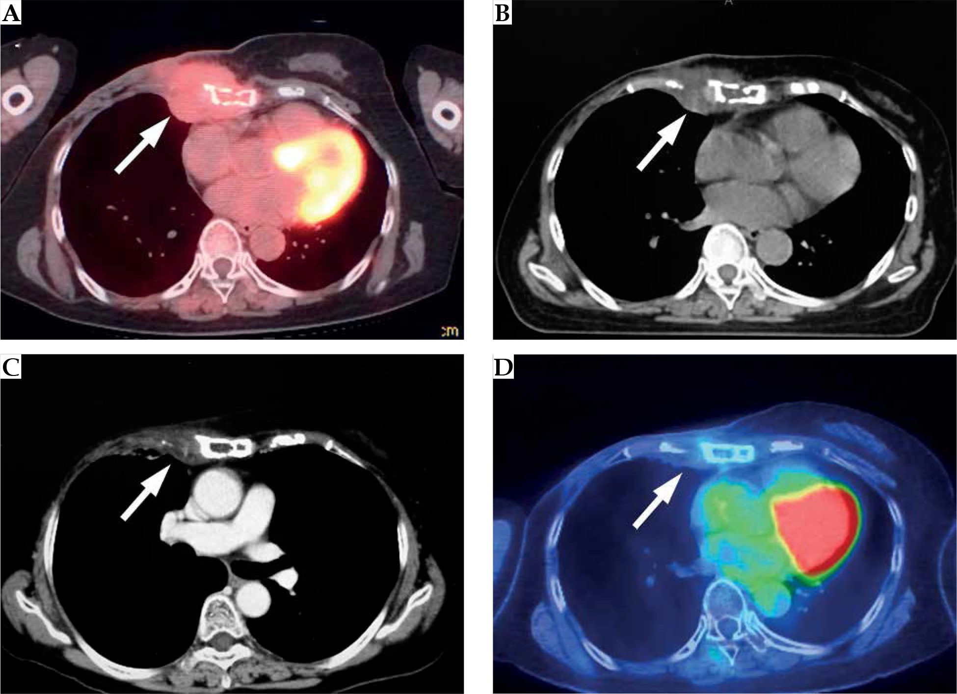
a) The white arrow indicates pericardial effusion, the blue arrow ...
Frontiers | Pathological complete response in a patient with pleural ...
[PDF] Low dose peripheral systemic thrombolysis for treatment of ...
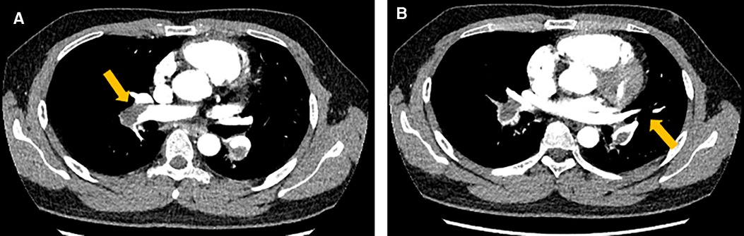
Three-dimensional CT aortography (axial view). (A-C) Aneurysmal change ...
Mediastinal mass – Radiology Cases
Lung cancer: squamous cell carcinoma – Radiology Cases
Computed tomography (CT) images in a case of severe post-thoracic ...
(PDF) Renal cell carcinoma with intramyocardial metastases
Faces of Bronchiectasis | Lungs
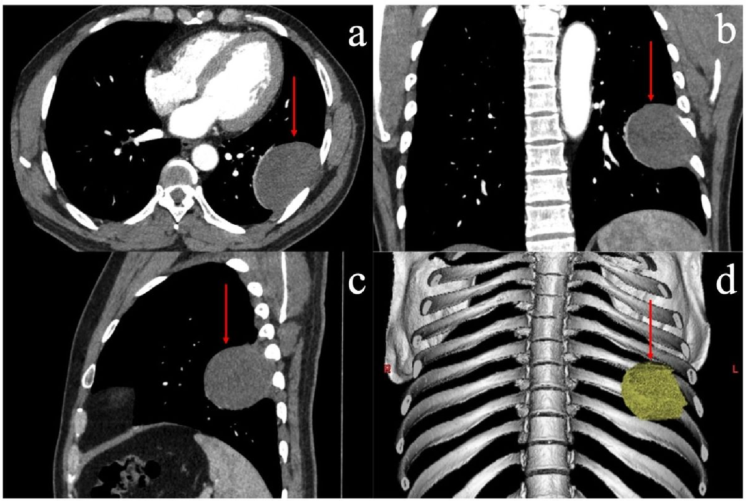
a) Axial contrast-enhanced CT scan. Heterogeneous well capsulated ...
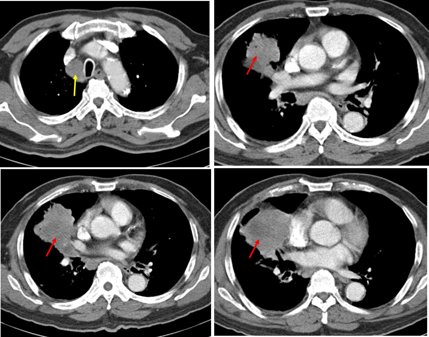
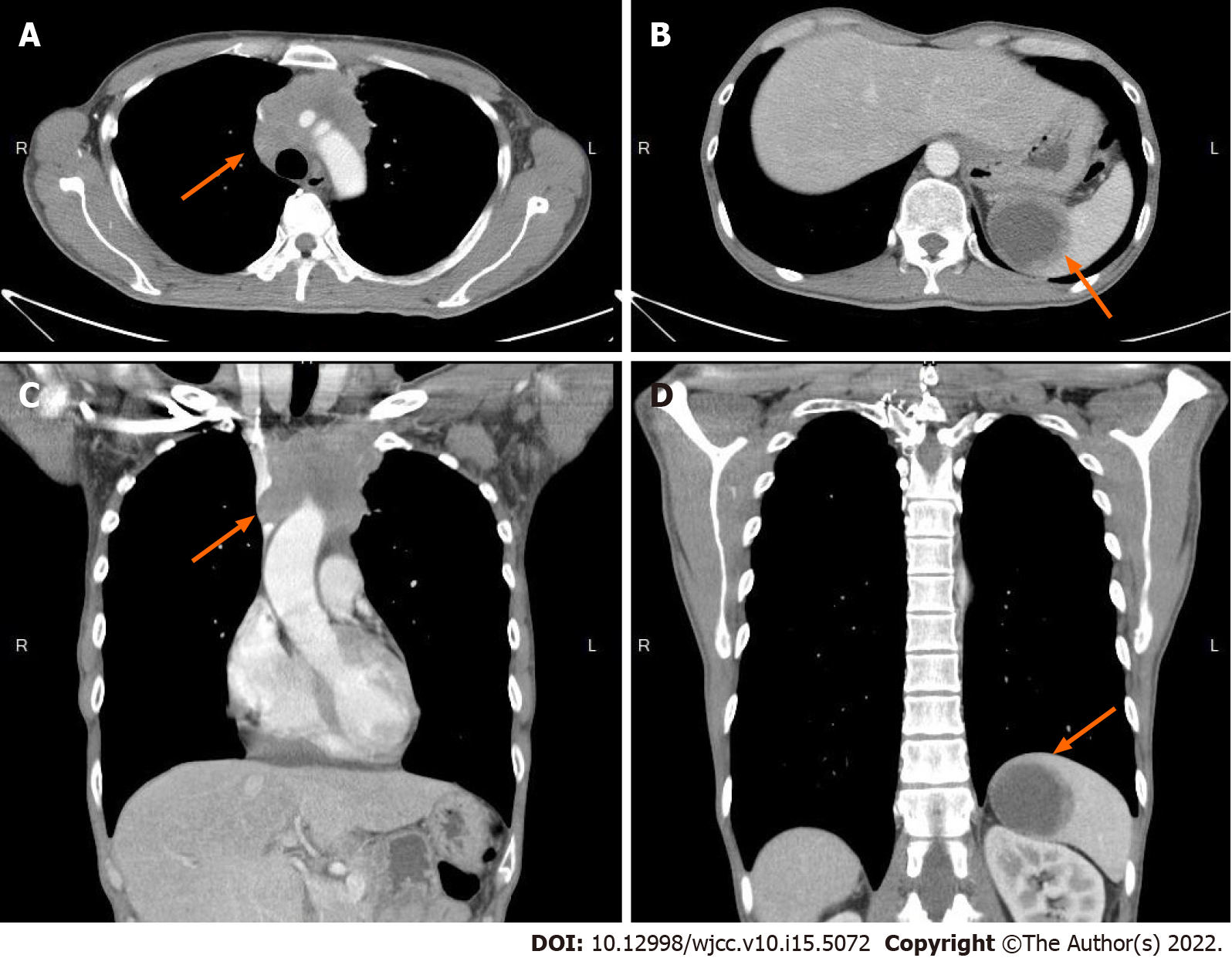
Chest CT scan. (A-D) Sequence of images in the mediastinal window ...
SVC Occlusion with Portal Vein Collaterals
“Vanishing” breast implant – when a breast prosthesis is moving into ...
Preoperative computed tomography shows a right mediastinal mass with ...
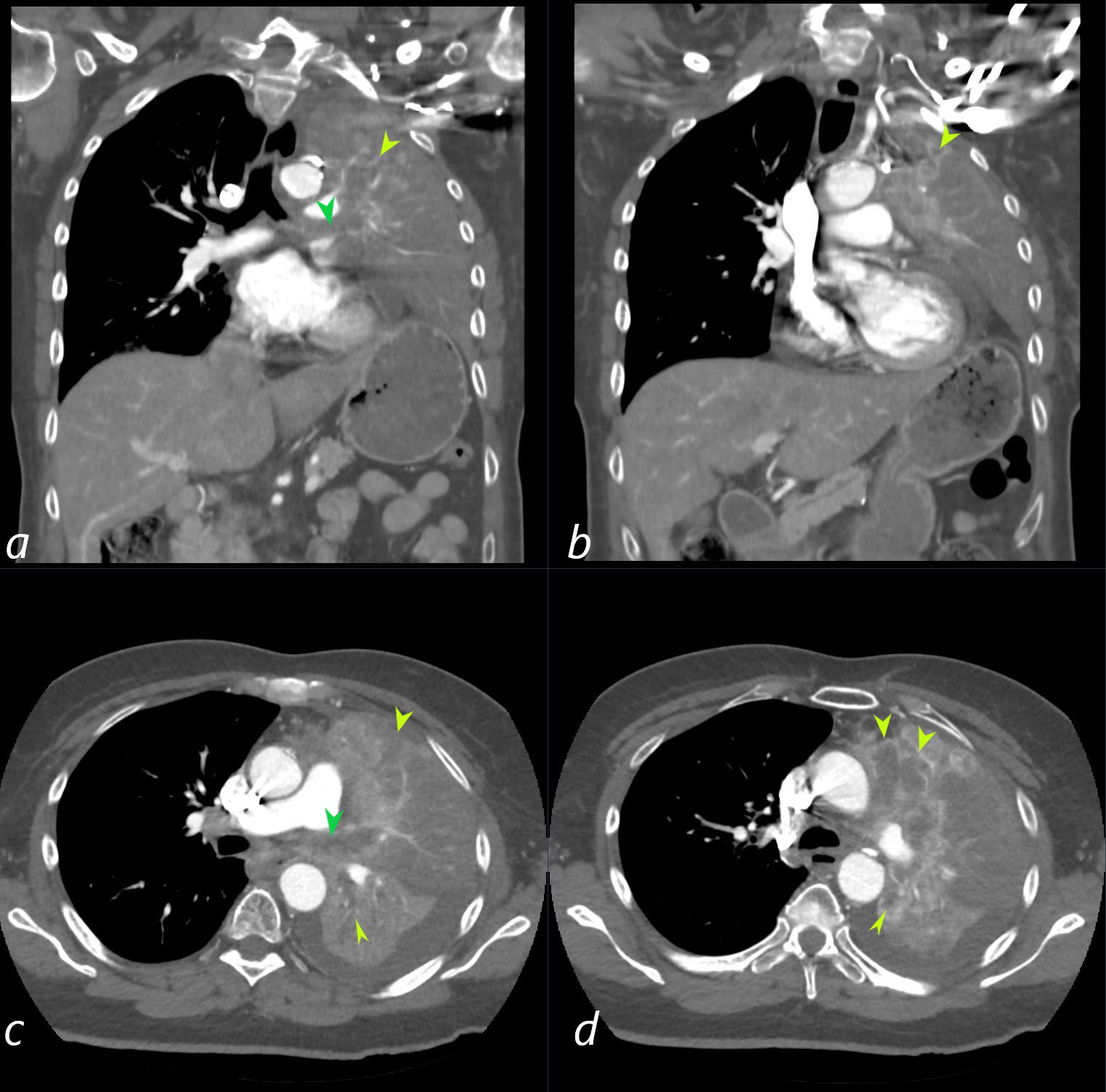
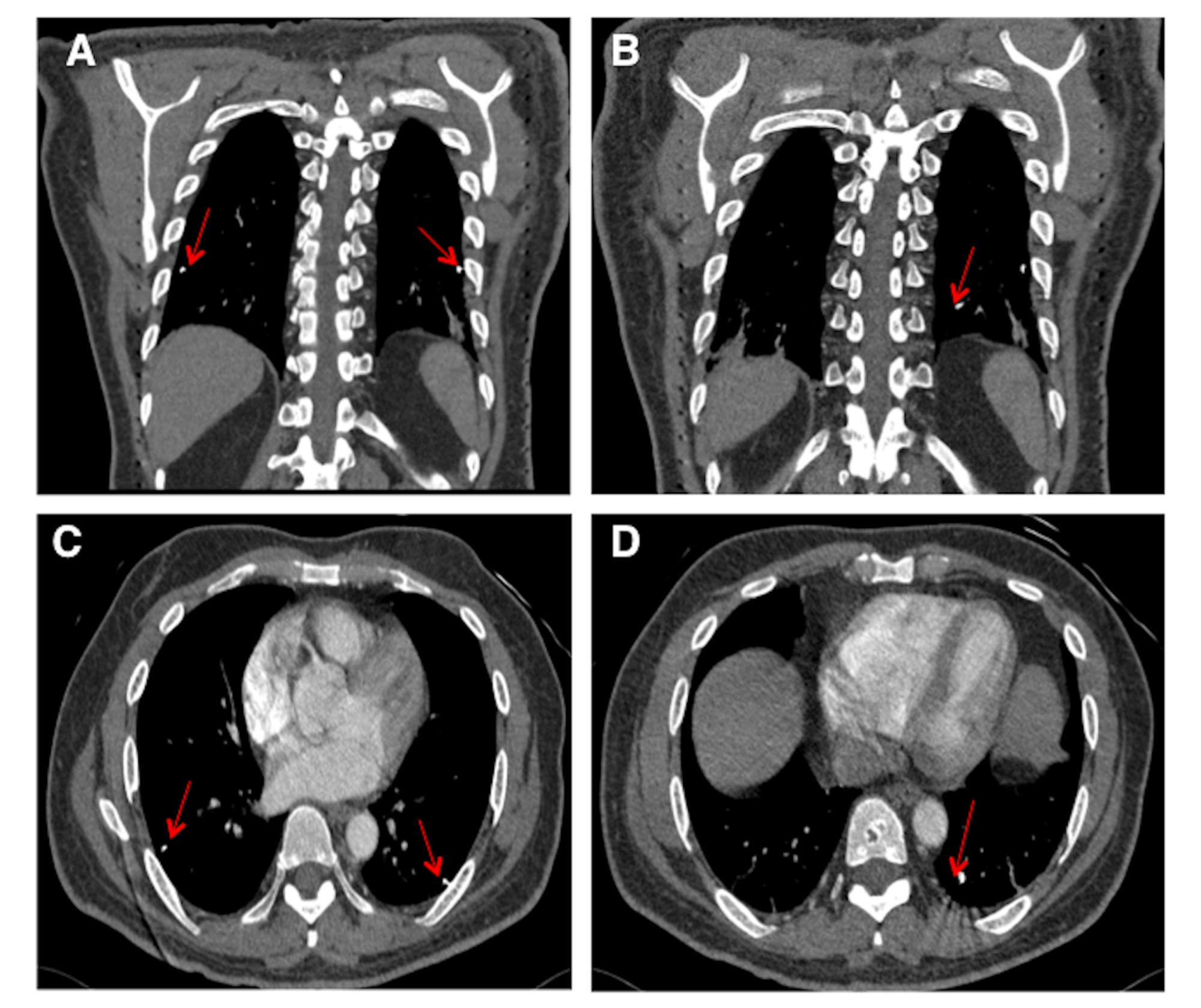
(a) Computed tomography (CT) pulmonary angiography axial plane shows ...
000 Asbestos and Diseases Caused by Asbestos | Lungs
Frontiers | Evaluation of CT features for differentiating consolidation ...
Rare solitary splenic metastasis from a thymic carcinoma detected on ...
Cureus | A Case Report of Foreign Body Embolization
Computed tomography images demonstrate a decrease in the size of lung ...
(a): Enhanced thoracic computed tomography: axial view: Heterogenous ...
Pre operative CT Pulmonary Angiography axial sections -: | Download ...
Unresectable bulky chest wall recurrent breast cancer controlled with ...
Mediastinal hemangiomas: Spectrum of CT and MRI findings ...
Saddle Pulmonary Embolism with RV Strain
Anomalous Pulmonary Venous Drainage and Pulmonary Vein Stenosis ...
Figure 2:Unusual Presentation Chronic Pulmonary Embolism due to ...
Preoperative imaging diagnoses. (A) A barium swallow identified an ...
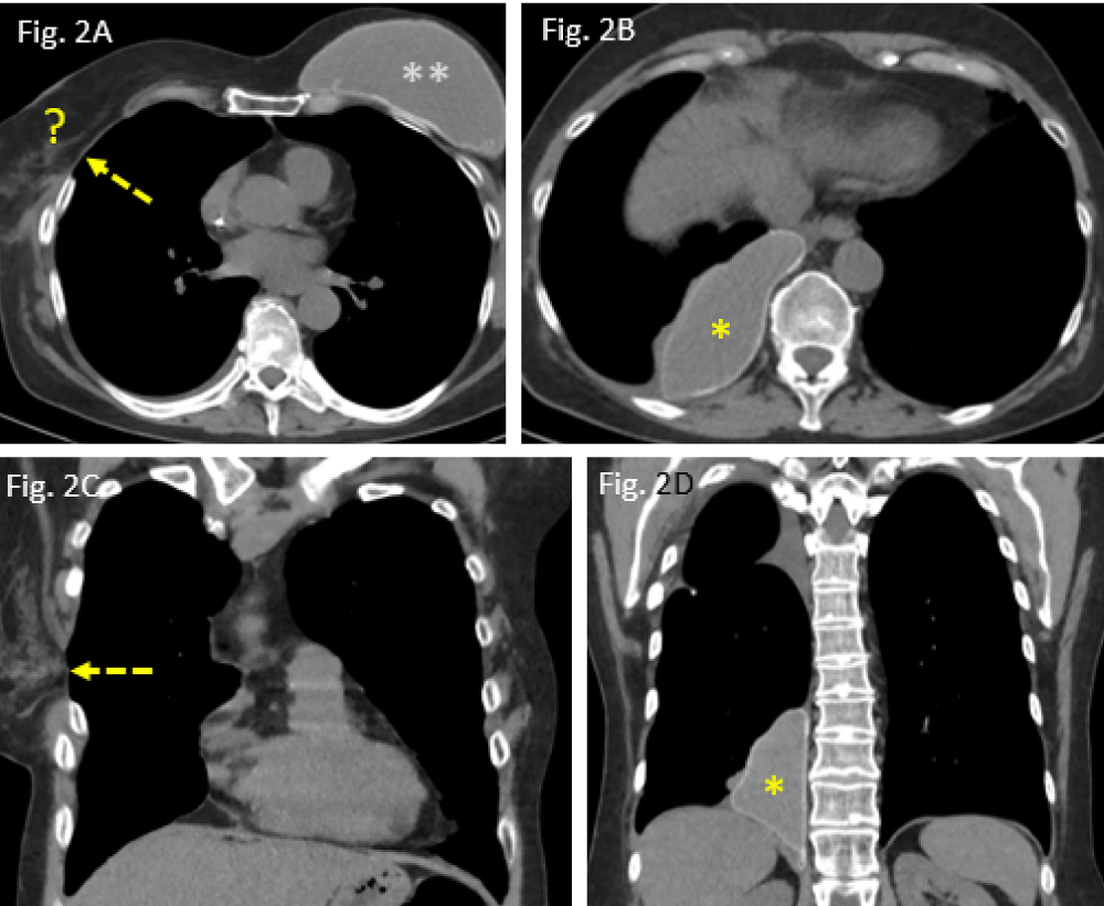
Chest axial CT (a) and sagittal (b) in the mediastinal window ...
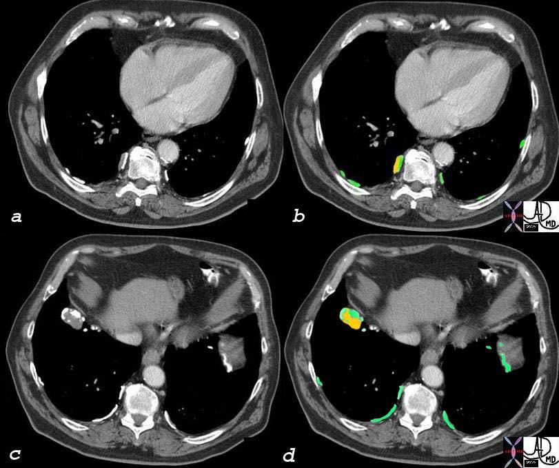
Iodine Distribution Map in Dual-Energy Computed Tomography Pulmonary ...
Frontiers | Myocarditis complicated by massive right ventricular ...
Three-dimensional CT aortography (axial view). (A-C) Aneurysmal change ...
Figure 1 from Schwannoma of Left Chest Wall: a Case Report and ...
Mediastinal Tuberculoma Mimicking Malignant Cardiac Tumor: A Case ...
Case of the Week Submission Information - American Osteopathic College ...


![[PDF] Low dose peripheral systemic thrombolysis for treatment of ...](https://d3i71xaburhd42.cloudfront.net/dc4ed39c976a1eddec5a43f560e31de2daf5c468/4-Figure1-1.png)