Please enter url.
Login
Logout
Please enter url.
Cervical Lymph Nodes Diagram
mungfali.com
source
Comments
-Axial, contrast-enhanced CT images (A and B) through the neck, above ...
Serial computerized tomography findings. (A) Multiple conglomerated ...
CT scan demonstrating retrosternal extension of thyroid mass in a ...
Patient with 5 cm retrosternal goiter below the thoracic inlet (total ...
(PDF) A case report: Giant cystic parathyroid adenoma presenting with ...
(PDF) Plaque Vulnerability in Internal Carotid Arteries with Positive ...
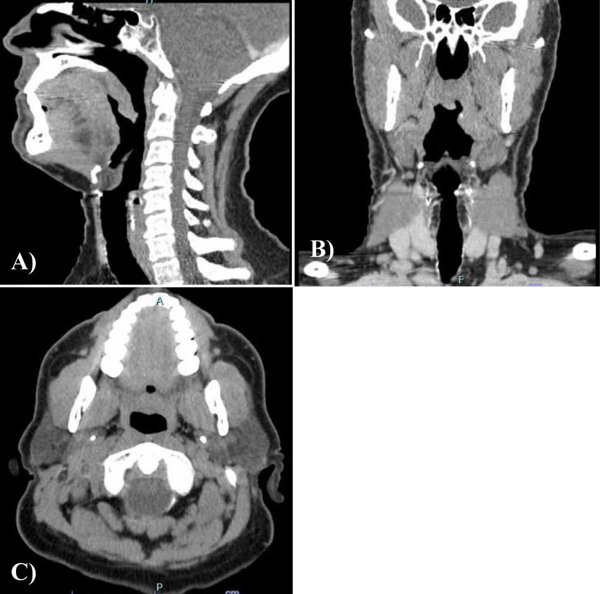
Radiologic manifestations of angioedema | SpringerLink
Coronal (A) and sagittal (B) views of CT angiogram of the chest and ...
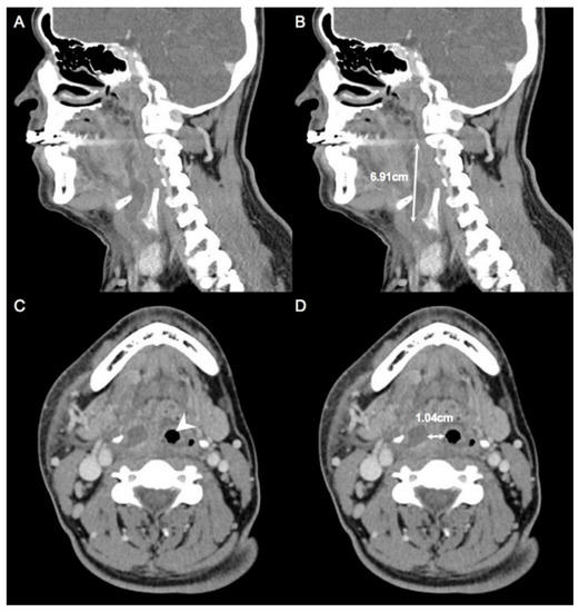
CT neck with contrast was performed. An approximately 1.2 cm ...
Case 1: pre-and post-treatment cervicothoracic CT; (A) pre-operative ...
Lung computed tomography: showing a soft tissue mass shadow in the ...
IGCCCG risk classification for advanced testicular cancer | Download Table
Figure: Gas in the retropharyngeal space (A) Plain cervical radiograph ...
(A) Repositioning of the axis on the PPV junction in OsiriX. (B ...
(PDF) Basaloid Squamous Cell Carcinoma, an Aggressive and Rare Cancer ...
Abstract 5788: VCAM-1 Targeted PET-CT Detects Inflammatory ...
CT scans of a 78-year-old female. CT scans revealing (A) an arc-shaped ...
Carcinoma Ex Pleomorphic Adenoma of the Uvula - Case Report
Diagnostics | Free Full-Text | Deep Learning Artificial Intelligence to ...
Subclavian (vertebral) steal syndrome | STROKE MANUAL
A 68-year-old male with angiotensin converting enzyme (ACE ...
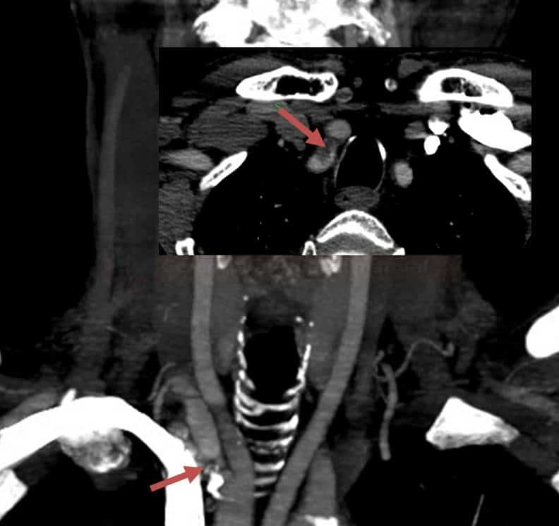
-(a) Axial CT angiogram obtained at the time of the patient's first ...
CT scan of the neck and paranasal sinuses. (A) Axial view showing the ...
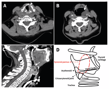
Laryngeal stridor in rheumatoid arthritis | CMAJ
Post traumatic facial artery pseudoaneurysm-A case report
Pre-operative CT cephalometry and functional outcomes after open ...
Combination of pembrolizumab and lenvatinib is a potential t ...
A: Preoperative MSCT image in axial view showing a mass (Blue Arrow ...
Intracordal fat assessment by 3-dimensional imaging after autologous ...
Computed tomography images showing an example of hypopharyngeal ...
Diagnostic and Management Value of Multi-Slice Computed Tomography in ...
-Axial T1W (A), T2W (B, C) sequences brain MRI of left cerebellar ...
CT scan of the neck with contrast showing evidence of faint enhancement ...
Revista Fronteras en Medicina
Sporadic bilateral carotid body paragangliomas | BMJ Case Reports