Please enter url.
Login
Logout
Please enter url.
Bilateral Globus Pallidus Internus Deep Brain Stimulation in ...
clinmedjournals.org
source
Comments
Chimeric Antigen Receptor T-Cell Therapy and Imaging Applications for ...
Cureus | Posterior Reversible Encephalopathy Syndrome With Hemorrhagic ...
Frontiers | Acute Necrotizing Encephalopathy of Childhood: A ...
Zellweger Syndrome - Magnetic Resonance - 78 Steps Health Journal
Patient 1. Images at presentation (A) and follow-up 5 (B-E) and 45 (F ...
MRI of the patient 1 at the age of 7 months, and 11 months. A) T2W, 7 ...
Preoperative brain MRI demonstrates a mass lesion located in the left ...
Figure 1 from Moyamoya disease with occlusion of bilateral vertebral ...
THE LEUKODYSTROPHIES | Neupsy Key
Status Marmoratus - Magnetic Resonance - 78 Steps Health
MR-Revealed Myelination in the Cerebral Corticospinal Tract as a Marker ...
Pseudo-insular glioma syndrome: illustrative cases in: Journal of ...
Very late-onset mitochondrial cytopathy featuring epilepsia partialis ...
Brain (A) T2WI (B) FLAIR (C) DIR (D) FLAIR and (E) DIR. FLAIR ...
Brain MRI made at the age of 13 months (images A, B, and C) and at the ...
Encephalitis and Thalamic Injury From Neuroinvasive West Nile Virus in ...
Neurosyphilis presenting with gummatous oculomotor nerve palsy ...
The Mammillary Bodies: A Review of Causes of Injury in Infants and ...
Lissencephaly: Update on diagnostics and clinical management - European ...
-MRI of a 15-month-old boy with HIBCH. Axial DWI (A), ADC (B), and ...
Figure 2 from Synthetic Mri of the Brain in a Clinical Setting ...
MR imaging findings of each patient. Axial T2-weighted image of Case 1 ...
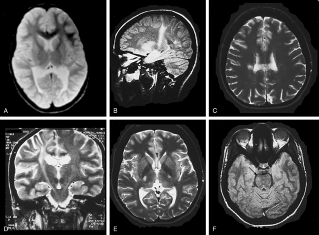
Clinical details and brain MRI from patient 4 with autoimmune ...
Occasional seizures, epilepsy, and inborn errors of metabolism - The ...
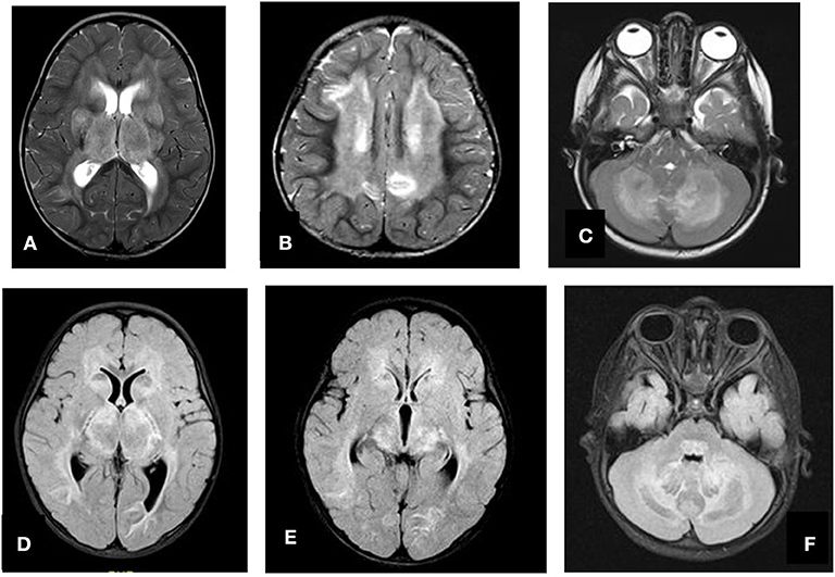
MRI of the brain (General Electric HDx Signa 3-T, USA) showing multiple ...
Cerebrale Hilus - Magnetic Resonance - 78 Steps Health Journal
Magnetic Resonance Imaging in Evaluation for Epilepsy Surgery | Neupsy Key
Diffusion tensor magnetic resonance imaging of glial brain tumors ...
Brain MRI imaging. T2-weighted TSE axial scans (1.5 T) at the level of ...
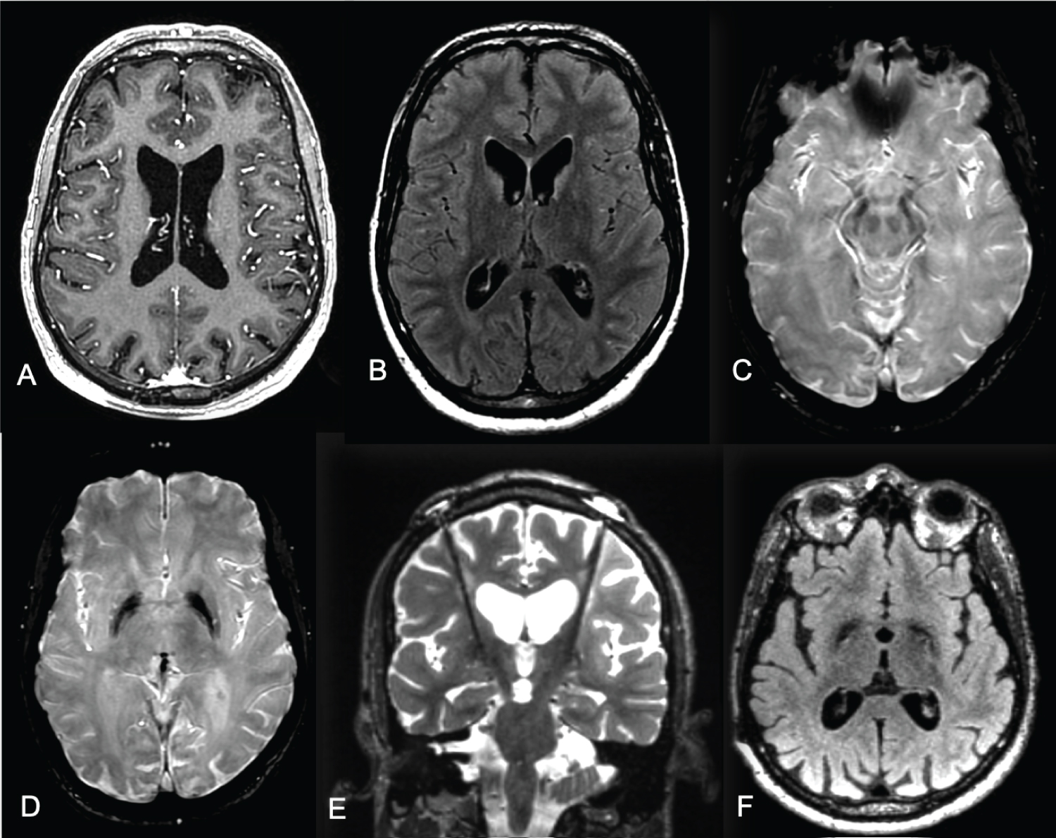
Imaging differences between ALSP and AARS2-L | Download Scientific Diagram
Brain MRI showed cortical thickening and loss of corticosubcortical ...
CADASIL is due to a NOTCH3 gene mutation. Although clinical symptoms ...
Magnetic resonance imaging in Case 4 show a right perisylvian mass ...
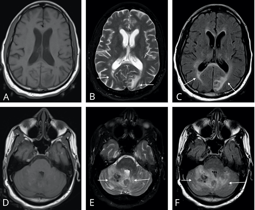
Brain MRI at the second admission. There is an extensive asymmetric ...
Lymphomatosis cerebri: a rare variant of primary central nervous system ...